Innovative Angiography System Supports Interventional Radiography
|
By MedImaging International staff writers Posted on 29 Mar 2017 |

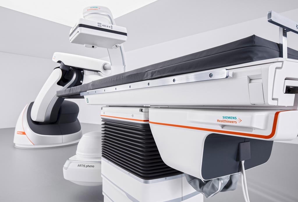
Image: The Artis pheno angiography system and tilt table (Photo courtesy of Siemens Healthineers).
An innovative robot-supported angiography system advances minimally invasive surgery, interventional radiology, and interventional cardiology in the hybrid operating room (OR).
The Siemens Healthineers Artis pheno angiography system is based on a the zen40HDR flat panel detector and the Gigalix X-ray tube, which provide the system with 2k imaging technology that offers a fourfold increase in resolution for 2D imaging, with up to 15% faster scan times in the body area. This increase is made possible by syngo DynaCT 3D imaging, which uses less contrast agent during the imaging process; if the patient is sensitive to contrast agents, Artis pheno can also support CO2 imaging of the extremities.
The C-arm has a free inner diameter of 95.5 centimeters, which offers more space for handling adipose and moribund patients, while the system’s multi-tilt table can accommodate patients weighing up to 280 kilograms. The robotic construction provides a flexible isocenter that allows the C-arm to follow all table positions and provide the best possible imaging support for the patient's treatment, presenting the target area of the body from virtually any angle. The end of the table can also be tilted in order to stabilize the patient's blood pressure or to make breathing easier.
Optional application packages are available to suit complex cases. For example, up to ten vertebrae can be represented in 3D using syngo DynaCT Large Volume, giving surgeons a larger overview during spinal fusion procedures. Another application, syngo DynaCT 360, can generate arge-volume images of the lung or the liver, including the anatomy of the tumor and the vessels leading to it, providing effective support for tumor transarterial chemoembolization (TACE), which involves supplying embolic particles coated with a chemotherapeutic drug via a catheter directly into the arteries leading to the tumor.
Another application, Syngo Needle Guidance, makes it possible to plan extensive procedures using orthopedic screws or surgical needles. Screw paths can be planned with precision, with an automatic path alignment function that automatically aligns the C-arm to follow them. A laser integrated in the image detector shows the surgeon the planned path, which helps improve both accuracy and speed in the OR and minimizes the rate of screw positioning errors in the spine.
Another new feature called StructureScout can adapt and optimize imaging parameters to best suit the material structure of the area being X-rayed, which enables even less radiation to be used, and also increases CO2 visibility when the table is tilted. The system also supports effective hygiene management in the hospital environment thanks to an antimicrobial coating, CleanGuide technology, and large sealed surfaces with fewer spaces, which helps customers with system cleaning.
“We see a high number of multi-morbid patients with impaired kidney function in the angio suite,” said Professor Frank Wacker, MD, director of the Institute for Diagnostic and Interventional Radiology at Hanover Medical School. “Shorter scan times help reduce the amount of iodinated contrast agent during 3D angiography in the thorax and abdomen by up to 15 percent.”
Latest Radiography News
- Novel Breast Imaging System Proves As Effective As Mammography
- AI Assistance Improves Breast-Cancer Screening by Reducing False Positives
- AI Could Boost Clinical Adoption of Chest DDR
- 3D Mammography Almost Halves Breast Cancer Incidence between Two Screening Tests
- AI Model Predicts 5-Year Breast Cancer Risk from Mammograms
- Deep Learning Framework Detects Fractures in X-Ray Images With 99% Accuracy
- Direct AI-Based Medical X-Ray Imaging System a Paradigm-Shift from Conventional DR and CT
- Chest X-Ray AI Solution Automatically Identifies, Categorizes and Highlights Suspicious Areas
- AI Diagnoses Wrist Fractures As Well As Radiologists
- Annual Mammography Beginning At 40 Cuts Breast Cancer Mortality By 42%
- 3D Human GPS Powered By Light Paves Way for Radiation-Free Minimally-Invasive Surgery
- Novel AI Technology to Revolutionize Cancer Detection in Dense Breasts
- AI Solution Provides Radiologists with 'Second Pair' Of Eyes to Detect Breast Cancers
- AI Helps General Radiologists Achieve Specialist-Level Performance in Interpreting Mammograms
- Novel Imaging Technique Could Transform Breast Cancer Detection
- Computer Program Combines AI and Heat-Imaging Technology for Early Breast Cancer Detection
Channels
MRI
view channel
World's First Whole-Body Ultra-High Field MRI Officially Comes To Market
The world's first whole-body ultra-high field (UHF) MRI has officially come to market, marking a remarkable advancement in diagnostic radiology. United Imaging (Shanghai, China) has secured clearance from the U.... Read more
World's First Sensor Detects Errors in MRI Scans Using Laser Light and Gas
MRI scanners are daily tools for doctors and healthcare professionals, providing unparalleled 3D imaging of the brain, vital organs, and soft tissues, far surpassing other imaging technologies in quality.... Read more
Diamond Dust Could Offer New Contrast Agent Option for Future MRI Scans
Gadolinium, a heavy metal used for over three decades as a contrast agent in medical imaging, enhances the clarity of MRI scans by highlighting affected areas. Despite its utility, gadolinium not only... Read more.jpg)
Combining MRI with PSA Testing Improves Clinical Outcomes for Prostate Cancer Patients
Prostate cancer is a leading health concern globally, consistently being one of the most common types of cancer among men and a major cause of cancer-related deaths. In the United States, it is the most... Read moreUltrasound
view channel
First AI-Powered POC Ultrasound Diagnostic Solution Helps Prioritize Cases Based On Severity
Ultrasound scans are essential for identifying and diagnosing various medical conditions, but often, patients must wait weeks or months for results due to a shortage of qualified medical professionals... Read more
Largest Model Trained On Echocardiography Images Assesses Heart Structure and Function
Foundation models represent an exciting frontier in generative artificial intelligence (AI), yet many lack the specialized medical data needed to make them applicable in healthcare settings.... Read more.jpg)
Groundbreaking Technology Enables Precise, Automatic Measurement of Peripheral Blood Vessels
The current standard of care of using angiographic information is often inadequate for accurately assessing vessel size in the estimated 20 million people in the U.S. who suffer from peripheral vascular disease.... Read moreNuclear Medicine
view channelNew PET Agent Rapidly and Accurately Visualizes Lesions in Clear Cell Renal Cell Carcinoma Patients
Clear cell renal cell cancer (ccRCC) represents 70-80% of renal cell carcinoma cases. While localized disease can be effectively treated with surgery and ablative therapies, one-third of patients either... Read more
New Imaging Technique Monitors Inflammation Disorders without Radiation Exposure
Imaging inflammation using traditional radiological techniques presents significant challenges, including radiation exposure, poor image quality, high costs, and invasive procedures. Now, new contrast... Read more
New SPECT/CT Technique Could Change Imaging Practices and Increase Patient Access
The development of lead-212 (212Pb)-PSMA–based targeted alpha therapy (TAT) is garnering significant interest in treating patients with metastatic castration-resistant prostate cancer. The imaging of 212Pb,... Read moreGeneral/Advanced Imaging
view channel
Radiation Therapy Computed Tomography Solution Boosts Imaging Accuracy
One of the most significant challenges in oncology care is disease complexity in terms of the variety of cancer types and the individualized presentation of each patient. This complexity necessitates a... Read more
PET Scans Reveal Hidden Inflammation in Multiple Sclerosis Patients
A key challenge for clinicians treating patients with multiple sclerosis (MS) is that after a certain amount of time, they continue to worsen even though their MRIs show no change. A new study has now... Read moreImaging IT
view channel
New Google Cloud Medical Imaging Suite Makes Imaging Healthcare Data More Accessible
Medical imaging is a critical tool used to diagnose patients, and there are billions of medical images scanned globally each year. Imaging data accounts for about 90% of all healthcare data1 and, until... Read more
Global AI in Medical Diagnostics Market to Be Driven by Demand for Image Recognition in Radiology
The global artificial intelligence (AI) in medical diagnostics market is expanding with early disease detection being one of its key applications and image recognition becoming a compelling consumer proposition... Read moreIndustry News
view channel
Hologic Acquires UK-Based Breast Surgical Guidance Company Endomagnetics Ltd.
Hologic, Inc. (Marlborough, MA, USA) has entered into a definitive agreement to acquire Endomagnetics Ltd. (Cambridge, UK), a privately held developer of breast cancer surgery technologies, for approximately... Read more
Bayer and Google Partner on New AI Product for Radiologists
Medical imaging data comprises around 90% of all healthcare data, and it is a highly complex and rich clinical data modality and serves as a vital tool for diagnosing patients. Each year, billions of medical... Read more


