Fear Over Low-Dose Imaging Radiation is Overstated
|
By MedImaging International staff writers Posted on 02 Feb 2017 |

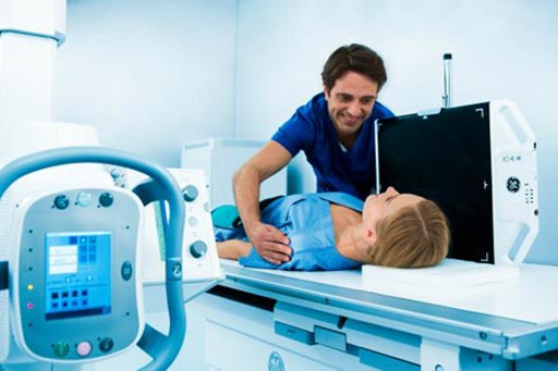
Image: New research shows diagnostic imaging radiation may not promote cancer (Photo courtesy of ITN).
The long-held belief that even low doses of radiation, such as those received in diagnostic imaging, increase cancer risk is based on an inaccurate, 70-year-old hypothesis, according to a new study.
Researchers at Nuclear Physics Enterprises (Marlton, NJ, USA) and the U.S. Food and Drug Administration argue that the claim that radiologic imaging carries an iatrogenic risk of cancer is based on an uninformed commitment to the 70-year-old linear no-threshold hypothesis (LNTH). The argument asserts that LNTH and its offspring, the low as reasonably achievable (ALARA) theory, are fatally flawed, focusing only on molecular damage while ignoring protective, organismal biologic responses.
The researchers show that credible evidence of imaging-related low-dose (i.e., lower than 100 mGy) carcinogenic risk is nonexistent, and it is a hypothetical risk derived from the demonstrably false LNTH. On the contrary, they claim, low doses of radiation stimulate protective responses, providing enhanced protection against additional damage over time, including damage from subsequent, higher radiation exposures. In essence, low-dose radiation does not cause, but more likely helps prevent cancer. The study was published in the January 2017 issue of The Journal of Nuclear Medicine.
“We have shown that the claim made by Hermann Muller during his 1946 Nobel Lecture that all radiation is harmful, regardless of how low the dose and dose rate was a non-sequitur unrecognized by the radiation science community,” said lead author Jeffry Siegel, PhD, president and CEO of Nuclear Physics Enterprises. “It has repeatedly been shown that the dose-response relationship may reasonably be considered to be linear but only down to a threshold, below which there is no demonstrable harm and even often benefit. Yet, the LNTH still rules radiation regulatory policy.”
“The task before us is to undo the public’s groundless fears of low-dose radiation exposure. The medical profession must be properly re-educated, beginning with diagnostic radiologists and nuclear medicine physicians, and only then can the public be given valid information that they can trust,” concluded Dr. Siegel. “Furthermore, defeating the LNTH and its offspring ALARA may lead to new ways of diagnosing and treating illness, and, even more importantly, preventing it.”
Data from the atomic-bomb survivor life span study (LSS) show the LNTH-predicted, low-dose carcinogenicity is invalid below approximately 200 mGy. The effective dose of a typical computed tomography (CT) scan is about 10 mSv; a positron emission tomography (PET) brain scan, 5-7 mSv; and a routine whole-body F-18 FDG PET/CT scan, 12-15 mSv. The researchers therefore feel confident that medical imaging's much lower doses for children or adults should not be feared or avoided for radio-phobic reasons.
Latest Radiography News
- Novel Breast Imaging System Proves As Effective As Mammography
- AI Assistance Improves Breast-Cancer Screening by Reducing False Positives
- AI Could Boost Clinical Adoption of Chest DDR
- 3D Mammography Almost Halves Breast Cancer Incidence between Two Screening Tests
- AI Model Predicts 5-Year Breast Cancer Risk from Mammograms
- Deep Learning Framework Detects Fractures in X-Ray Images With 99% Accuracy
- Direct AI-Based Medical X-Ray Imaging System a Paradigm-Shift from Conventional DR and CT
- Chest X-Ray AI Solution Automatically Identifies, Categorizes and Highlights Suspicious Areas
- AI Diagnoses Wrist Fractures As Well As Radiologists
- Annual Mammography Beginning At 40 Cuts Breast Cancer Mortality By 42%
- 3D Human GPS Powered By Light Paves Way for Radiation-Free Minimally-Invasive Surgery
- Novel AI Technology to Revolutionize Cancer Detection in Dense Breasts
- AI Solution Provides Radiologists with 'Second Pair' Of Eyes to Detect Breast Cancers
- AI Helps General Radiologists Achieve Specialist-Level Performance in Interpreting Mammograms
- Novel Imaging Technique Could Transform Breast Cancer Detection
- Computer Program Combines AI and Heat-Imaging Technology for Early Breast Cancer Detection
Channels
MRI
view channel
Diamond Dust Could Offer New Contrast Agent Option for Future MRI Scans
Gadolinium, a heavy metal used for over three decades as a contrast agent in medical imaging, enhances the clarity of MRI scans by highlighting affected areas. Despite its utility, gadolinium not only... Read more.jpg)
Combining MRI with PSA Testing Improves Clinical Outcomes for Prostate Cancer Patients
Prostate cancer is a leading health concern globally, consistently being one of the most common types of cancer among men and a major cause of cancer-related deaths. In the United States, it is the most... Read more
PET/MRI Improves Diagnostic Accuracy for Prostate Cancer Patients
The Prostate Imaging Reporting and Data System (PI-RADS) is a five-point scale to assess potential prostate cancer in MR images. PI-RADS category 3 which offers an unclear suggestion of clinically significant... Read more
Next Generation MR-Guided Focused Ultrasound Ushers In Future of Incisionless Neurosurgery
Essential tremor, often called familial, idiopathic, or benign tremor, leads to uncontrollable shaking that significantly affects a person’s life. When traditional medications do not alleviate symptoms,... Read moreUltrasound
view channel.jpg)
Groundbreaking Technology Enables Precise, Automatic Measurement of Peripheral Blood Vessels
The current standard of care of using angiographic information is often inadequate for accurately assessing vessel size in the estimated 20 million people in the U.S. who suffer from peripheral vascular disease.... Read more
Deep Learning Advances Super-Resolution Ultrasound Imaging
Ultrasound localization microscopy (ULM) is an advanced imaging technique that offers high-resolution visualization of microvascular structures. It employs microbubbles, FDA-approved contrast agents, injected... Read more
Novel Ultrasound-Launched Targeted Nanoparticle Eliminates Biofilm and Bacterial Infection
Biofilms, formed by bacteria aggregating into dense communities for protection against harsh environmental conditions, are a significant contributor to various infectious diseases. Biofilms frequently... Read moreNuclear Medicine
view channel
New Imaging Technique Monitors Inflammation Disorders without Radiation Exposure
Imaging inflammation using traditional radiological techniques presents significant challenges, including radiation exposure, poor image quality, high costs, and invasive procedures. Now, new contrast... Read more
New SPECT/CT Technique Could Change Imaging Practices and Increase Patient Access
The development of lead-212 (212Pb)-PSMA–based targeted alpha therapy (TAT) is garnering significant interest in treating patients with metastatic castration-resistant prostate cancer. The imaging of 212Pb,... Read moreNew Radiotheranostic System Detects and Treats Ovarian Cancer Noninvasively
Ovarian cancer is the most lethal gynecological cancer, with less than a 30% five-year survival rate for those diagnosed in late stages. Despite surgery and platinum-based chemotherapy being the standard... Read more
AI System Automatically and Reliably Detects Cardiac Amyloidosis Using Scintigraphy Imaging
Cardiac amyloidosis, a condition characterized by the buildup of abnormal protein deposits (amyloids) in the heart muscle, severely affects heart function and can lead to heart failure or death without... Read moreGeneral/Advanced Imaging
view channel
PET Scans Reveal Hidden Inflammation in Multiple Sclerosis Patients
A key challenge for clinicians treating patients with multiple sclerosis (MS) is that after a certain amount of time, they continue to worsen even though their MRIs show no change. A new study has now... Read more
Artificial Intelligence Evaluates Cardiovascular Risk from CT Scans
Chest computed tomography (CT) is a common diagnostic tool, with approximately 15 million scans conducted each year in the United States, though many are underutilized or not fully explored.... Read more
New AI Method Captures Uncertainty in Medical Images
In the field of biomedicine, segmentation is the process of annotating pixels from an important structure in medical images, such as organs or cells. Artificial Intelligence (AI) models are utilized to... Read more.jpg)
CT Coronary Angiography Reduces Need for Invasive Tests to Diagnose Coronary Artery Disease
Coronary artery disease (CAD), one of the leading causes of death worldwide, involves the narrowing of coronary arteries due to atherosclerosis, resulting in insufficient blood flow to the heart muscle.... Read moreImaging IT
view channel
New Google Cloud Medical Imaging Suite Makes Imaging Healthcare Data More Accessible
Medical imaging is a critical tool used to diagnose patients, and there are billions of medical images scanned globally each year. Imaging data accounts for about 90% of all healthcare data1 and, until... Read more
Global AI in Medical Diagnostics Market to Be Driven by Demand for Image Recognition in Radiology
The global artificial intelligence (AI) in medical diagnostics market is expanding with early disease detection being one of its key applications and image recognition becoming a compelling consumer proposition... Read moreIndustry News
view channel
Bayer and Google Partner on New AI Product for Radiologists
Medical imaging data comprises around 90% of all healthcare data, and it is a highly complex and rich clinical data modality and serves as a vital tool for diagnosing patients. Each year, billions of medical... Read more


