Deep-Learning Algorithm Detects Vertebral Compression Fractures
|
By MedImaging International staff writers Posted on 01 Feb 2017 |

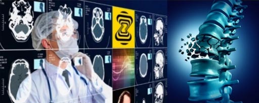
Image: A novel algorithm helps detect vertical compression fractures (Photo courtesy of Zebra Medical Vision).
A novel algorithm can differentiate between a vertical compression fracture (VCF) and more ubiquitous degenerative endplate changes and osteophytes.
The Zebra Medical Vision VCF algorithm automatically segments the vertebral column in order to identify and localize compression fractures. Diagnosing VCF’s is of critical importance for implementation of both primary (therapeutic) and secondary (preventative) osteoporotic interventions. As such, implementation of the algorithm can help prevent a large number of VCFs, allowing for better preventative and overall care, as well as reducing long term healthcare costs for providers.
The algorithm was developed utilizing a combination of traditional machine vision segmentation and convolutional neural net (CNN) technology, and can be applied to any computerized tomography (CT) scan of the chest, abdomen, and/or pelvis. As such, it will become the latest addition to a line of automated tools announced by Zebra Medical Vision, among them algorithms that automatically detect low bone mineral density, breast cancer, fatty liver, coronary artery calcium, emphysema, and more.
“Radiology is headed for a personnel crisis for a number of reasons: the increase in population, especially the elderly and ill, increased exposure of the developing countries to radiology services, and the increase in the quantity of information from imaging devices, while the number of radiologists has not changed,” said Elad Benjamin, co-founder and CEO of Zebra Medical Vision. “We want to help radiologists analyze the images, while saving time, and to free them, so that they can devote their efforts to the more complex cases.”
“Research has shown that radiologists miss up to 50% of vertebral fractures, since they are usually focused on looking for other features,” added Kassim Javiad, MD, of Oxford University Hospitals (United Kingdom). “In the UK, with our proven coordinated care programs for effective fracture prevention, we believe that early detection of such fractures can yield both better care and significant healthcare cost savings.”
VCFs are a direct cause of morbidity, decreasing mobility and functional status particularly among the elderly. Osteoporotic VCFs affect up to one in four of post -menopausal women and nearly one in seven men over the age of 65. Timely surgical or minimally invasive treatment of VCF’s is effective but under-utilized, in part because less than one third of VCF’s are effectively diagnosed. Although VCF’s may be the result of infection, trauma or malignancy, the vast majority are a manifestation of osteoporosis, especially in individuals over the age of 50.
Latest General/Advanced Imaging News
- Bone Density Test Uses Existing CT Images to Predict Fractures
- AI Predicts Cardiac Risk and Mortality from Routine Chest CT Scans
- Radiation Therapy Computed Tomography Solution Boosts Imaging Accuracy
- PET Scans Reveal Hidden Inflammation in Multiple Sclerosis Patients
- Artificial Intelligence Evaluates Cardiovascular Risk from CT Scans
- New AI Method Captures Uncertainty in Medical Images
- CT Coronary Angiography Reduces Need for Invasive Tests to Diagnose Coronary Artery Disease
- Novel Blood Test Could Reduce Need for PET Imaging of Patients with Alzheimer’s
- CT-Based Deep Learning Algorithm Accurately Differentiates Benign From Malignant Vertebral Fractures
- Minimally Invasive Procedure Could Help Patients Avoid Thyroid Surgery
- Self-Driving Mobile C-Arm Reduces Imaging Time during Surgery
- AR Application Turns Medical Scans Into Holograms for Assistance in Surgical Planning
- Imaging Technology Provides Ground-Breaking New Approach for Diagnosing and Treating Bowel Cancer
- CT Coronary Calcium Scoring Predicts Heart Attacks and Strokes
- AI Model Detects 90% of Lymphatic Cancer Cases from PET and CT Images
- Breakthrough Technology Revolutionizes Breast Imaging
Channels
Radiography
view channel
Novel Breast Imaging System Proves As Effective As Mammography
Breast cancer remains the most frequently diagnosed cancer among women. It is projected that one in eight women will be diagnosed with breast cancer during her lifetime, and one in 42 women who turn 50... Read more
AI Assistance Improves Breast-Cancer Screening by Reducing False Positives
Radiologists typically detect one case of cancer for every 200 mammograms reviewed. However, these evaluations often result in false positives, leading to unnecessary patient recalls for additional testing,... Read moreMRI
view channel
Low-Cost Whole-Body MRI Device Combined with AI Generates High-Quality Results
Magnetic Resonance Imaging (MRI) has significantly transformed healthcare, providing a noninvasive, radiation-free method for detailed imaging. It is especially promising for the future of medical diagnosis... Read more
World's First Whole-Body Ultra-High Field MRI Officially Comes To Market
The world's first whole-body ultra-high field (UHF) MRI has officially come to market, marking a remarkable advancement in diagnostic radiology. United Imaging (Shanghai, China) has secured clearance from the U.... Read moreUltrasound
view channel.jpg)
Diagnostic System Automatically Analyzes TTE Images to Identify Congenital Heart Disease
Congenital heart disease (CHD) is one of the most prevalent congenital anomalies worldwide, presenting substantial health and financial challenges for affected patients. Early detection and treatment of... Read more
Super-Resolution Imaging Technique Could Improve Evaluation of Cardiac Conditions
The heart depends on efficient blood circulation to pump blood throughout the body, delivering oxygen to tissues and removing carbon dioxide and waste. Yet, when heart vessels are damaged, it can disrupt... Read more
First AI-Powered POC Ultrasound Diagnostic Solution Helps Prioritize Cases Based On Severity
Ultrasound scans are essential for identifying and diagnosing various medical conditions, but often, patients must wait weeks or months for results due to a shortage of qualified medical professionals... Read moreNuclear Medicine
view channelNew PET Agent Rapidly and Accurately Visualizes Lesions in Clear Cell Renal Cell Carcinoma Patients
Clear cell renal cell cancer (ccRCC) represents 70-80% of renal cell carcinoma cases. While localized disease can be effectively treated with surgery and ablative therapies, one-third of patients either... Read more
New Imaging Technique Monitors Inflammation Disorders without Radiation Exposure
Imaging inflammation using traditional radiological techniques presents significant challenges, including radiation exposure, poor image quality, high costs, and invasive procedures. Now, new contrast... Read more
New SPECT/CT Technique Could Change Imaging Practices and Increase Patient Access
The development of lead-212 (212Pb)-PSMA–based targeted alpha therapy (TAT) is garnering significant interest in treating patients with metastatic castration-resistant prostate cancer. The imaging of 212Pb,... Read moreImaging IT
view channel
New Google Cloud Medical Imaging Suite Makes Imaging Healthcare Data More Accessible
Medical imaging is a critical tool used to diagnose patients, and there are billions of medical images scanned globally each year. Imaging data accounts for about 90% of all healthcare data1 and, until... Read more
Global AI in Medical Diagnostics Market to Be Driven by Demand for Image Recognition in Radiology
The global artificial intelligence (AI) in medical diagnostics market is expanding with early disease detection being one of its key applications and image recognition becoming a compelling consumer proposition... Read moreIndustry News
view channel
Hologic Acquires UK-Based Breast Surgical Guidance Company Endomagnetics Ltd.
Hologic, Inc. (Marlborough, MA, USA) has entered into a definitive agreement to acquire Endomagnetics Ltd. (Cambridge, UK), a privately held developer of breast cancer surgery technologies, for approximately... Read more
Bayer and Google Partner on New AI Product for Radiologists
Medical imaging data comprises around 90% of all healthcare data, and it is a highly complex and rich clinical data modality and serves as a vital tool for diagnosing patients. Each year, billions of medical... Read more


