Total Body Digital Skin Imaging System Developed for Dermatology and Primary Care Practices
|
By MedImaging International staff writers Posted on 03 Dec 2014 |

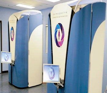
Image: The DermSpectra total body digital skin imaging system (Photo courtesy of DermSpectra).
A total body digital skin imaging system enables physicians to track critical skin changes (skin cancers, eczema, lesions, psoriasis, and rashes) in their office, over time.
The DermSpectra (Tucson, AZ, USA) medical technology advances the way physicians digitally capture and compare skin irregularities by rapidly delivering patented high-resolution imaging, HIPPA (Health Insurance Portability and Accountability Act of 1996)-compliant secure storage, and immediate viewing capabilities.
For use by patients, dermatology and primary care practices, telemedicine centers, cosmetic and plastic surgeons, and the medical community, DermSpectra fulfills a crucial gap in the digital skin imaging (DSI) market. Currently, there are few tools for standardized screening to aid in early detection of skin cancer at the total body level.
Using the system, patients experience an in-office private, convenient, and short imaging session (8–10 minutes) while their physician views the latest digital images (on desktop or iPad), annotates directly to the image, and stores them securely on a HIPPA-compliant server database. DermSpectra total body digital skin imaging enables physicians to complete the physical exam, assessment, and plan care during one office visit, lessening post-appointment documentation. The Apple (Cupertino, CA, USA) Store recently approved the DermSpectra viewing application, which is now available by physicians to view stored images.
Digital images captured through DermSpectra can be marked using simple touch screen features that allow physicians to zoom in on lesions, circle abnormalities or changes, add notes, annotate directly on the image, and attach a report to the patient’s electronic medical record (EMR). Ultimately this will move the field of dermatological assessment into an imagecentric-based practice.
According to Dr. Richard Carmona, 17th US Surgeon General and DermSpectra board member, “DermSpectra is setting the industry standard for how future dermatology, primary care, and medical practices will capture image data to assist early detection of melanoma and other skin diseases. With skin cancer incidence and treatment costs on the rise, DermSpectra is filling an imaging gap to aid in preventative skin screening.”
“DermSpectra total body digital skin imaging has been in beta testing since September 2013, most recently at Southwest Skin Specialists in Scottsdale, Arizona [USA] and University of Arizona Cancer Center in Tucson, Arizona, and is scheduled for installation at Mayo Clinic,” stated Karleen Seybold, co-founder and chief executive officer of DermSpectra. “Successful beta tests showed that 86% of participants reported being very satisfied with their DermSpectra experience and 88% said total body imaging is very important to the quality of medical care. DermSpectra is changing the way the industry and public thinks about preventative skin imaging and will standardize the way skin changes are tracked and monitored over time.”
The DermSpectra total body digital skin imaging system is a proprietary solution that provides patented high-resolution imaging, secure storage, and immediate viewing capabilities.
Related Links:
DermSpectra
Latest General/Advanced Imaging News
- AI Predicts Cardiac Risk and Mortality from Routine Chest CT Scans
- Radiation Therapy Computed Tomography Solution Boosts Imaging Accuracy
- PET Scans Reveal Hidden Inflammation in Multiple Sclerosis Patients
- Artificial Intelligence Evaluates Cardiovascular Risk from CT Scans
- New AI Method Captures Uncertainty in Medical Images
- CT Coronary Angiography Reduces Need for Invasive Tests to Diagnose Coronary Artery Disease
- Novel Blood Test Could Reduce Need for PET Imaging of Patients with Alzheimer’s
- CT-Based Deep Learning Algorithm Accurately Differentiates Benign From Malignant Vertebral Fractures
- Minimally Invasive Procedure Could Help Patients Avoid Thyroid Surgery
- Self-Driving Mobile C-Arm Reduces Imaging Time during Surgery
- AR Application Turns Medical Scans Into Holograms for Assistance in Surgical Planning
- Imaging Technology Provides Ground-Breaking New Approach for Diagnosing and Treating Bowel Cancer
- CT Coronary Calcium Scoring Predicts Heart Attacks and Strokes
- AI Model Detects 90% of Lymphatic Cancer Cases from PET and CT Images
- Breakthrough Technology Revolutionizes Breast Imaging
- State-Of-The-Art System Enhances Accuracy of Image-Guided Diagnostic and Interventional Procedures
Channels
Radiography
view channel
Novel Breast Imaging System Proves As Effective As Mammography
Breast cancer remains the most frequently diagnosed cancer among women. It is projected that one in eight women will be diagnosed with breast cancer during her lifetime, and one in 42 women who turn 50... Read more
AI Assistance Improves Breast-Cancer Screening by Reducing False Positives
Radiologists typically detect one case of cancer for every 200 mammograms reviewed. However, these evaluations often result in false positives, leading to unnecessary patient recalls for additional testing,... Read moreMRI
view channel
Low-Cost Whole-Body MRI Device Combined with AI Generates High-Quality Results
Magnetic Resonance Imaging (MRI) has significantly transformed healthcare, providing a noninvasive, radiation-free method for detailed imaging. It is especially promising for the future of medical diagnosis... Read more
World's First Whole-Body Ultra-High Field MRI Officially Comes To Market
The world's first whole-body ultra-high field (UHF) MRI has officially come to market, marking a remarkable advancement in diagnostic radiology. United Imaging (Shanghai, China) has secured clearance from the U.... Read moreUltrasound
view channel.jpg)
Diagnostic System Automatically Analyzes TTE Images to Identify Congenital Heart Disease
Congenital heart disease (CHD) is one of the most prevalent congenital anomalies worldwide, presenting substantial health and financial challenges for affected patients. Early detection and treatment of... Read more
Super-Resolution Imaging Technique Could Improve Evaluation of Cardiac Conditions
The heart depends on efficient blood circulation to pump blood throughout the body, delivering oxygen to tissues and removing carbon dioxide and waste. Yet, when heart vessels are damaged, it can disrupt... Read more
First AI-Powered POC Ultrasound Diagnostic Solution Helps Prioritize Cases Based On Severity
Ultrasound scans are essential for identifying and diagnosing various medical conditions, but often, patients must wait weeks or months for results due to a shortage of qualified medical professionals... Read moreNuclear Medicine
view channelNew PET Agent Rapidly and Accurately Visualizes Lesions in Clear Cell Renal Cell Carcinoma Patients
Clear cell renal cell cancer (ccRCC) represents 70-80% of renal cell carcinoma cases. While localized disease can be effectively treated with surgery and ablative therapies, one-third of patients either... Read more
New Imaging Technique Monitors Inflammation Disorders without Radiation Exposure
Imaging inflammation using traditional radiological techniques presents significant challenges, including radiation exposure, poor image quality, high costs, and invasive procedures. Now, new contrast... Read more
New SPECT/CT Technique Could Change Imaging Practices and Increase Patient Access
The development of lead-212 (212Pb)-PSMA–based targeted alpha therapy (TAT) is garnering significant interest in treating patients with metastatic castration-resistant prostate cancer. The imaging of 212Pb,... Read moreImaging IT
view channel
New Google Cloud Medical Imaging Suite Makes Imaging Healthcare Data More Accessible
Medical imaging is a critical tool used to diagnose patients, and there are billions of medical images scanned globally each year. Imaging data accounts for about 90% of all healthcare data1 and, until... Read more
Global AI in Medical Diagnostics Market to Be Driven by Demand for Image Recognition in Radiology
The global artificial intelligence (AI) in medical diagnostics market is expanding with early disease detection being one of its key applications and image recognition becoming a compelling consumer proposition... Read moreIndustry News
view channel
Hologic Acquires UK-Based Breast Surgical Guidance Company Endomagnetics Ltd.
Hologic, Inc. (Marlborough, MA, USA) has entered into a definitive agreement to acquire Endomagnetics Ltd. (Cambridge, UK), a privately held developer of breast cancer surgery technologies, for approximately... Read more
Bayer and Google Partner on New AI Product for Radiologists
Medical imaging data comprises around 90% of all healthcare data, and it is a highly complex and rich clinical data modality and serves as a vital tool for diagnosing patients. Each year, billions of medical... Read more


