Risk of Breast Cancer Later in Life for Girls Undergoing Radiotherapy Explained
|
By MedImaging International staff writers Posted on 02 Oct 2013 |

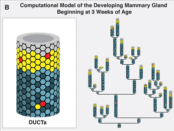
Image: Computation model of the developing mammary gland beginning at three weeks of age (Photo courtesy of the US Department of Energy’s Lawrence Berkeley National Lab’s Life Sciences Division
Exposing young women and girls under the age of 20 to ionizing radiation can substantially raise the risk of their developing breast cancer later in life. Scientists may now have found the answer as to why this occurs. A collaborative study’s findings pointed to increased stem cell self-renewal and consequential mammary stem cell enrichment as the reason.
Breasts enriched with mammary stem cells as a result of ionizing irradiation during puberty show a later-in-life propensity for developing estrogen receptor (ER)-negative tumors. Estrogen receptors, i.e., proteins activated by the estrogen hormone, are vital for the normal development of the breast and other female sexual features during puberty.
“Our results are in agreement with epidemiology studies showing that radiation-induced human breast cancers are more likely to be ER-negative than are spontaneous breast cancers,” said Dr. Sylvain Costes, a biophysicist with US Department of Energy’s (DOE) Lawrence Berkeley National Lab’s Life Sciences Division (LBL; Berkeley, CA, USA). “This is important because ER-negative breast cancers are less differentiated, more aggressive, and often have a poor prognosis compared to the other breast cancer subtypes.”
Drs. Costes and Jonathan Tang, also with Berkeley Lab’s Life Sciences Division, were part of a collaboration led by Dr. Mary Helen Barcellos-Hoff, formerly with Berkeley Lab and now at the New York University School of Medicine (New York, NY, USA), that studied the so-called “window of susceptibility” known to exist between radiation treatments at puberty and breast cancer risk in later adulthood. The key to their success were two mammary lineage agent-based models (ABMs) they developed in which a system is modeled as a collection of autonomous decision-making units called agents. One ABM simulated the effects of radiation on the mammary gland during either the developmental stages or during adulthood. The other simulated the growth dynamics of human mammary epithelial cells in culture after irradiation.
“Our mammary gland ABM consisted of millions of agents, with each agent representing either a mammary stem cell, a progenitor cell or a differentiated cell in the breast,” stated Dr. Tang. “We ran thousands of simulations on Berkeley Lab’s Lawrencium supercomputer during which each agent continually assessed its situation and made decisions on the basis of a set of rules that correspond to known or hypothesized biological properties of mammary cells. The advantage of this approach is that it allows us to view the global consequences to the system that emerge over time from our assumptions about the individual agents. To our knowledge, our mammary gland model is the first multiscale model of the development of full glands starting from the onset of puberty all the way to adulthood.”
Epidemiologic research have shown that girls under 20 administered radiotherapy treatment for disorders such as Hodgkin’s lymphoma have about the same risk of developing breast cancer in their 40s as women who were born with a BRCA gene mutation. From their study, Drs. Costes, Tang, and their colleagues concluded that self-renewal of stem cells was the most likely responsible process.
“Stem cell self-renewal was the only mechanism in the mammary gland model that led to predictions that were consistent with data from both our in vivo mouse work and our in vitro experiments with MCF10A, a human mammary epithelial cell line,” Dr. Tang noted. “Additionally, our model predicts that this mechanism would only generate more stem cells during puberty while the gland is developing and considerable cell proliferation is taking place.”
The investigators are now looking for genetic or phenotypic biomarkers that would identify young girls who are at the greatest breast cancer risk from radiotherapy. The study’s findings from Dr. Barcellos-Hoff and her research group show that the ties between ionizing radiation and breast cancer extend beyond DNA damage and mutations.
“Essentially, exposure of the breast to ionizing radiation generates an overall biochemical signal that tells the system something bad happened,” Dr. Costes said. “If exposure takes place during puberty, this signal triggers a regenerative response leading to a larger pool of stem cells, thereby increasing the chance of developing aggressive ER-negative breast cancers later in life.”
The findings of this collaborative study have been published online August 23, 2013, in the journal Stem Cells.
Related Links:
Lawrence Berkeley US National Lab’s Life Sciences Division
Latest Radiography News
- Novel Breast Imaging System Proves As Effective As Mammography
- AI Assistance Improves Breast-Cancer Screening by Reducing False Positives
- AI Could Boost Clinical Adoption of Chest DDR
- 3D Mammography Almost Halves Breast Cancer Incidence between Two Screening Tests
- AI Model Predicts 5-Year Breast Cancer Risk from Mammograms
- Deep Learning Framework Detects Fractures in X-Ray Images With 99% Accuracy
- Direct AI-Based Medical X-Ray Imaging System a Paradigm-Shift from Conventional DR and CT
- Chest X-Ray AI Solution Automatically Identifies, Categorizes and Highlights Suspicious Areas
- AI Diagnoses Wrist Fractures As Well As Radiologists
- Annual Mammography Beginning At 40 Cuts Breast Cancer Mortality By 42%
- 3D Human GPS Powered By Light Paves Way for Radiation-Free Minimally-Invasive Surgery
- Novel AI Technology to Revolutionize Cancer Detection in Dense Breasts
- AI Solution Provides Radiologists with 'Second Pair' Of Eyes to Detect Breast Cancers
- AI Helps General Radiologists Achieve Specialist-Level Performance in Interpreting Mammograms
- Novel Imaging Technique Could Transform Breast Cancer Detection
- Computer Program Combines AI and Heat-Imaging Technology for Early Breast Cancer Detection
Channels
MRI
view channel
Diamond Dust Could Offer New Contrast Agent Option for Future MRI Scans
Gadolinium, a heavy metal used for over three decades as a contrast agent in medical imaging, enhances the clarity of MRI scans by highlighting affected areas. Despite its utility, gadolinium not only... Read more.jpg)
Combining MRI with PSA Testing Improves Clinical Outcomes for Prostate Cancer Patients
Prostate cancer is a leading health concern globally, consistently being one of the most common types of cancer among men and a major cause of cancer-related deaths. In the United States, it is the most... Read more
PET/MRI Improves Diagnostic Accuracy for Prostate Cancer Patients
The Prostate Imaging Reporting and Data System (PI-RADS) is a five-point scale to assess potential prostate cancer in MR images. PI-RADS category 3 which offers an unclear suggestion of clinically significant... Read more
Next Generation MR-Guided Focused Ultrasound Ushers In Future of Incisionless Neurosurgery
Essential tremor, often called familial, idiopathic, or benign tremor, leads to uncontrollable shaking that significantly affects a person’s life. When traditional medications do not alleviate symptoms,... Read moreUltrasound
view channel.jpg)
Groundbreaking Technology Enables Precise, Automatic Measurement of Peripheral Blood Vessels
The current standard of care of using angiographic information is often inadequate for accurately assessing vessel size in the estimated 20 million people in the U.S. who suffer from peripheral vascular disease.... Read more
Deep Learning Advances Super-Resolution Ultrasound Imaging
Ultrasound localization microscopy (ULM) is an advanced imaging technique that offers high-resolution visualization of microvascular structures. It employs microbubbles, FDA-approved contrast agents, injected... Read more
Novel Ultrasound-Launched Targeted Nanoparticle Eliminates Biofilm and Bacterial Infection
Biofilms, formed by bacteria aggregating into dense communities for protection against harsh environmental conditions, are a significant contributor to various infectious diseases. Biofilms frequently... Read moreNuclear Medicine
view channel
New Imaging Technique Monitors Inflammation Disorders without Radiation Exposure
Imaging inflammation using traditional radiological techniques presents significant challenges, including radiation exposure, poor image quality, high costs, and invasive procedures. Now, new contrast... Read more
New SPECT/CT Technique Could Change Imaging Practices and Increase Patient Access
The development of lead-212 (212Pb)-PSMA–based targeted alpha therapy (TAT) is garnering significant interest in treating patients with metastatic castration-resistant prostate cancer. The imaging of 212Pb,... Read moreNew Radiotheranostic System Detects and Treats Ovarian Cancer Noninvasively
Ovarian cancer is the most lethal gynecological cancer, with less than a 30% five-year survival rate for those diagnosed in late stages. Despite surgery and platinum-based chemotherapy being the standard... Read more
AI System Automatically and Reliably Detects Cardiac Amyloidosis Using Scintigraphy Imaging
Cardiac amyloidosis, a condition characterized by the buildup of abnormal protein deposits (amyloids) in the heart muscle, severely affects heart function and can lead to heart failure or death without... Read moreGeneral/Advanced Imaging
view channel
PET Scans Reveal Hidden Inflammation in Multiple Sclerosis Patients
A key challenge for clinicians treating patients with multiple sclerosis (MS) is that after a certain amount of time, they continue to worsen even though their MRIs show no change. A new study has now... Read more
Artificial Intelligence Evaluates Cardiovascular Risk from CT Scans
Chest computed tomography (CT) is a common diagnostic tool, with approximately 15 million scans conducted each year in the United States, though many are underutilized or not fully explored.... Read more
New AI Method Captures Uncertainty in Medical Images
In the field of biomedicine, segmentation is the process of annotating pixels from an important structure in medical images, such as organs or cells. Artificial Intelligence (AI) models are utilized to... Read more.jpg)
CT Coronary Angiography Reduces Need for Invasive Tests to Diagnose Coronary Artery Disease
Coronary artery disease (CAD), one of the leading causes of death worldwide, involves the narrowing of coronary arteries due to atherosclerosis, resulting in insufficient blood flow to the heart muscle.... Read moreImaging IT
view channel
New Google Cloud Medical Imaging Suite Makes Imaging Healthcare Data More Accessible
Medical imaging is a critical tool used to diagnose patients, and there are billions of medical images scanned globally each year. Imaging data accounts for about 90% of all healthcare data1 and, until... Read more
Global AI in Medical Diagnostics Market to Be Driven by Demand for Image Recognition in Radiology
The global artificial intelligence (AI) in medical diagnostics market is expanding with early disease detection being one of its key applications and image recognition becoming a compelling consumer proposition... Read moreIndustry News
view channel
Bayer and Google Partner on New AI Product for Radiologists
Medical imaging data comprises around 90% of all healthcare data, and it is a highly complex and rich clinical data modality and serves as a vital tool for diagnosing patients. Each year, billions of medical... Read more


