AI Helps Optimize CT Scan X-Ray Radiation Dose
|
By MedImaging International staff writers Posted on 10 Mar 2023 |

Computed tomography (CT) is a highly effective and extensive diagnostic tool utilized by modern medicine. Unfortunately, there is growing concern regarding the increasing number of patients undergoing CT scans, and the considerable amount of X-ray radiation to which they are exposed. The ALARA principle, commonly known as "As Low As Reasonably Achievable," implies that a patient should receive the most significant diagnostic benefit with minimal radiation exposure. In practical terms, this principle requires a trade-off since decreasing the level of radiation administered typically results in poorer CT image quality. Accordingly, medical professionals must strike a balance between obtaining high-quality CT images and minimizing a patient's exposure to X-rays to reduce the risk of misdiagnosis.
To strike a balance between image quality and radiation exposure during CT scans, healthcare professionals, including radiologists, may employ an optimization strategy. First, they observe actual images generated by the tomographer to identify abnormalities such as tumors or unusual tissue. Statistical methods are then utilized to calculate the optimal radiation dose and tomographer configuration. This procedure can be generalized by adopting reference CT images obtained by scanning specially designed phantoms that comprise inserts of varying sizes and contrasts, which represent standardized abnormalities. However, manual image analysis is incredibly time-consuming. To address this issue, a team of researchers from the University of Florence (Florence, Italy), in collaboration with radiologists and medical physicists, examined if this process could be automated by using artificial intelligence (AI). The team created and trained an algorithm - a “model observer” - based on convolutional neural networks (CNNs), which could analyze the standardized abnormalities in CT images as efficiently as a professional.
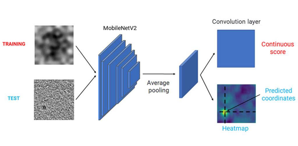
The team needed sufficient training and testing data for their model, for which 30 healthcare professionals visually examined 1000 CT images in a phantom mimicking human tissue. The phantom contained cylindrical inserts of varying diameters and contrasts, and the observers had to identify whether an object was present in the image as well as indicate the level of confidence in their assessment. This generated a dataset of 30,000 labeled CT images captured using various tomographic reconstruction configurations, accurately reflecting human interpretation. The team then implemented two AI models based on different architectures - UNet and MobileNetV2 – and modified the base design of these architectures to allow them perform both classification ("Is there an unusual object in the CT image?") and localization ("Where is the unusual object?"). The models were then trained and tested using images from the dataset.
The research team conducted statistical analyses to evaluate various performance metrics to ensure that the model observers accurately emulated how a human would assess CT images of the phantom. The researchers are optimistic that with further efforts, their model can become a viable mechanism for automated CT image quality assessment. They are confident that applying their AI model observers on a larger scale will enable faster and safer CT evaluations than ever before.
“Our results were very promising, as both trained models performed remarkably well and achieved an absolute percentage error of less than 5%,” said Dr. Sandra Doria of the Physics Department at the University of Florence who led the research team. “This indicated that the models could identify the object inserted in the phantom with similar accuracy and confidence as a human professional, for almost all reconstruction configurations and abnormalities sizes and contrasts.”
Related Links:
University of Florence
Latest Radiography News
- World's Largest Class Single Crystal Diamond Radiation Detector Opens New Possibilities for Diagnostic Imaging
- AI-Powered Imaging Technique Shows Promise in Evaluating Patients for PCI
- Higher Chest X-Ray Usage Catches Lung Cancer Earlier and Improves Survival
- AI-Powered Mammograms Predict Cardiovascular Risk
- Generative AI Model Significantly Reduces Chest X-Ray Reading Time
- AI-Powered Mammography Screening Boosts Cancer Detection in Single-Reader Settings
- Photon Counting Detectors Promise Fast Color X-Ray Images
- AI Can Flag Mammograms for Supplemental MRI
- 3D CT Imaging from Single X-Ray Projection Reduces Radiation Exposure
- AI Method Accurately Predicts Breast Cancer Risk by Analyzing Multiple Mammograms
- Printable Organic X-Ray Sensors Could Transform Treatment for Cancer Patients
- Highly Sensitive, Foldable Detector to Make X-Rays Safer
- Novel Breast Cancer Screening Technology Could Offer Superior Alternative to Mammogram
- Artificial Intelligence Accurately Predicts Breast Cancer Years Before Diagnosis
- AI-Powered Chest X-Ray Detects Pulmonary Nodules Three Years Before Lung Cancer Symptoms
- AI Model Identifies Vertebral Compression Fractures in Chest Radiographs
Channels
Radiography
view channel
World's Largest Class Single Crystal Diamond Radiation Detector Opens New Possibilities for Diagnostic Imaging
Diamonds possess ideal physical properties for radiation detection, such as exceptional thermal and chemical stability along with a quick response time. Made of carbon with an atomic number of six, diamonds... Read more
AI-Powered Imaging Technique Shows Promise in Evaluating Patients for PCI
Percutaneous coronary intervention (PCI), also known as coronary angioplasty, is a minimally invasive procedure where small metal tubes called stents are inserted into partially blocked coronary arteries... Read moreMRI
view channel
AI Tool Tracks Effectiveness of Multiple Sclerosis Treatments Using Brain MRI Scans
Multiple sclerosis (MS) is a condition in which the immune system attacks the brain and spinal cord, leading to impairments in movement, sensation, and cognition. Magnetic Resonance Imaging (MRI) markers... Read more
Ultra-Powerful MRI Scans Enable Life-Changing Surgery in Treatment-Resistant Epileptic Patients
Approximately 360,000 individuals in the UK suffer from focal epilepsy, a condition in which seizures spread from one part of the brain. Around a third of these patients experience persistent seizures... Read more
AI-Powered MRI Technology Improves Parkinson’s Diagnoses
Current research shows that the accuracy of diagnosing Parkinson’s disease typically ranges from 55% to 78% within the first five years of assessment. This is partly due to the similarities shared by Parkinson’s... Read more
Biparametric MRI Combined with AI Enhances Detection of Clinically Significant Prostate Cancer
Artificial intelligence (AI) technologies are transforming the way medical images are analyzed, offering unprecedented capabilities in quantitatively extracting features that go beyond traditional visual... Read moreUltrasound
view channel
AI Identifies Heart Valve Disease from Common Imaging Test
Tricuspid regurgitation is a condition where the heart's tricuspid valve does not close completely during contraction, leading to backward blood flow, which can result in heart failure. A new artificial... Read more
Novel Imaging Method Enables Early Diagnosis and Treatment Monitoring of Type 2 Diabetes
Type 2 diabetes is recognized as an autoimmune inflammatory disease, where chronic inflammation leads to alterations in pancreatic islet microvasculature, a key factor in β-cell dysfunction.... Read moreNuclear Medicine
view channel
Novel PET Imaging Approach Offers Never-Before-Seen View of Neuroinflammation
COX-2, an enzyme that plays a key role in brain inflammation, can be significantly upregulated by inflammatory stimuli and neuroexcitation. Researchers suggest that COX-2 density in the brain could serve... Read more
Novel Radiotracer Identifies Biomarker for Triple-Negative Breast Cancer
Triple-negative breast cancer (TNBC), which represents 15-20% of all breast cancer cases, is one of the most aggressive subtypes, with a five-year survival rate of about 40%. Due to its significant heterogeneity... Read moreImaging IT
view channel
New Google Cloud Medical Imaging Suite Makes Imaging Healthcare Data More Accessible
Medical imaging is a critical tool used to diagnose patients, and there are billions of medical images scanned globally each year. Imaging data accounts for about 90% of all healthcare data1 and, until... Read more
Global AI in Medical Diagnostics Market to Be Driven by Demand for Image Recognition in Radiology
The global artificial intelligence (AI) in medical diagnostics market is expanding with early disease detection being one of its key applications and image recognition becoming a compelling consumer proposition... Read moreIndustry News
view channel
GE HealthCare and NVIDIA Collaboration to Reimagine Diagnostic Imaging
GE HealthCare (Chicago, IL, USA) has entered into a collaboration with NVIDIA (Santa Clara, CA, USA), expanding the existing relationship between the two companies to focus on pioneering innovation in... Read more
Patient-Specific 3D-Printed Phantoms Transform CT Imaging
New research has highlighted how anatomically precise, patient-specific 3D-printed phantoms are proving to be scalable, cost-effective, and efficient tools in the development of new CT scan algorithms... Read more
Siemens and Sectra Collaborate on Enhancing Radiology Workflows
Siemens Healthineers (Forchheim, Germany) and Sectra (Linköping, Sweden) have entered into a collaboration aimed at enhancing radiologists' diagnostic capabilities and, in turn, improving patient care... Read more


 Guided Devices.jpg)


