Antibacterial ‘Smart Stitches’ Reveal Location of Sutured Area in CT Scans
|
By MedImaging International staff writers Posted on 10 Feb 2023 |
Surgical site infections are among the most common medical infections and occur in 2 to 4% of patients following surgery, although the infection rate can be much higher for some procedures, such as vaginal mesh implants to treat prolapse, infection. Now, a new antimicrobial suture material that glows in medical imaging offers a promising alternative for mesh implants and internal stitches.
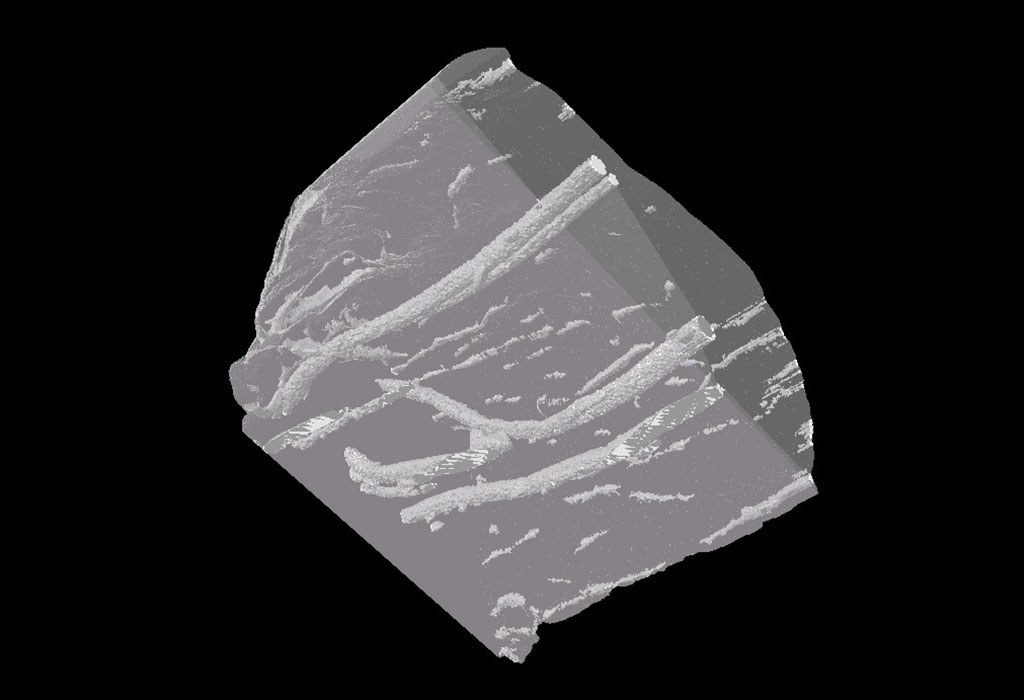
A multidisciplinary research team led by RMIT University (Melbourne, Australia) that included nano-engineering, biomedical and textile experts working in partnership with a practicing surgeon has developed the proof-of-concept material. In lab tests conducted by the team, the surgical filament threaded through samples of chicken meat was easily visible in CT scans, even after three weeks. The surgical filament also demonstrated strong antimicrobial properties by killing 99% of highly drug-resistant bacteria after six hours at body temperature.
The suture gains its properties from the combination of iodine and tiny nanoparticles, called carbon dots, throughout the material. Carbon dots are inherently fluorescent, due to their particular wavelength, but can also be tuned to different levels of luminosity that easily stand out from surrounding tissue in medical imaging. By attaching iodine to these carbon dots, they gain strong antimicrobial properties and greater X-ray visibility.
According to the researchers, carbon nano dots are safe, cheap and easy to produce in the lab from natural ingredients. The researchers believe that it addresses a serious challenge faced by surgeons in attempting to identify the precise anatomical location of internal meshes on CT scans. The researchers will now conduct pre-clinical trials for which they will produce larger suture samples.
“Our smart surgical sutures can play an important role in preventing infection and monitoring patient recovery and the proof-of-concept material we’ve developed has several important properties that make it an exciting candidate for this,” said Dr. Shadi Houshyar, study lead author and Vice Chancellor’s Senior Research Fellow from RMIT University’s School of Engineering.
“This mesh will enable us to help with improved identification of the causes of symptoms, reduce the incidence of mesh infections and will help with precise preoperative planning, if there is a need to surgically remove this mesh,” said consultant colorectal surgeon and Professor of Surgery at the University of Melbourne, Justin Yeung, who was involved in the study. “It has the potential to improve surgery outcomes and improve quality of life for a huge proportion of women, if used as vaginal mesh for example, by reducing the need for infected mesh removal. It may also significantly reduce surgery duration and increase surgical accuracy in general through the ability to visualize mesh location accurately on preoperative imaging.”
Related Links:
RMIT University
Latest Surgical Techniques News
Channels
Radiography
view channel
AI Algorithm Uses Mammograms to Accurately Predict Cardiovascular Risk in Women
Cardiovascular disease remains the leading cause of death in women worldwide, responsible for about nine million deaths annually. Despite this burden, symptoms and risk factors are often under-recognized... Read more
AI Hybrid Strategy Improves Mammogram Interpretation
Breast cancer screening programs rely heavily on radiologists interpreting mammograms, a process that is time-intensive and subject to errors. While artificial intelligence (AI) models have shown strong... Read moreMRI
view channel
AI-Assisted Model Enhances MRI Heart Scans
A cardiac MRI can reveal critical information about the heart’s function and any abnormalities, but traditional scans take 30 to 90 minutes and often suffer from poor image quality due to patient movement.... Read more
AI Model Outperforms Doctors at Identifying Patients Most At-Risk of Cardiac Arrest
Hypertrophic cardiomyopathy is one of the most common inherited heart conditions and a leading cause of sudden cardiac death in young individuals and athletes. While many patients live normal lives, some... Read moreUltrasound
view channel
Non-Invasive Ultrasound-Based Tool Accurately Detects Infant Meningitis
Meningitis, an inflammation of the membranes surrounding the brain and spinal cord, can be fatal in infants if not diagnosed and treated early. Even when treated, it may leave lasting damage, such as cognitive... Read more
Breakthrough Deep Learning Model Enhances Handheld 3D Medical Imaging
Ultrasound imaging is a vital diagnostic technique used to visualize internal organs and tissues in real time and to guide procedures such as biopsies and injections. When paired with photoacoustic imaging... Read moreNuclear Medicine
view channel
New Camera Sees Inside Human Body for Enhanced Scanning and Diagnosis
Nuclear medicine scans like single-photon emission computed tomography (SPECT) allow doctors to observe heart function, track blood flow, and detect hidden diseases. However, current detectors are either... Read more
Novel Bacteria-Specific PET Imaging Approach Detects Hard-To-Diagnose Lung Infections
Mycobacteroides abscessus is a rapidly growing mycobacteria that primarily affects immunocompromised patients and those with underlying lung diseases, such as cystic fibrosis or chronic obstructive pulmonary... Read moreGeneral/Advanced Imaging
view channel
New Ultrasmall, Light-Sensitive Nanoparticles Could Serve as Contrast Agents
Medical imaging technologies face ongoing challenges in capturing accurate, detailed views of internal processes, especially in conditions like cancer, where tracking disease development and treatment... Read more
AI Algorithm Accurately Predicts Pancreatic Cancer Metastasis Using Routine CT Images
In pancreatic cancer, detecting whether the disease has spread to other organs is critical for determining whether surgery is appropriate. If metastasis is present, surgery is not recommended, yet current... Read moreImaging IT
view channel
New Google Cloud Medical Imaging Suite Makes Imaging Healthcare Data More Accessible
Medical imaging is a critical tool used to diagnose patients, and there are billions of medical images scanned globally each year. Imaging data accounts for about 90% of all healthcare data1 and, until... Read more
Global AI in Medical Diagnostics Market to Be Driven by Demand for Image Recognition in Radiology
The global artificial intelligence (AI) in medical diagnostics market is expanding with early disease detection being one of its key applications and image recognition becoming a compelling consumer proposition... Read moreIndustry News
view channel
GE HealthCare and NVIDIA Collaboration to Reimagine Diagnostic Imaging
GE HealthCare (Chicago, IL, USA) has entered into a collaboration with NVIDIA (Santa Clara, CA, USA), expanding the existing relationship between the two companies to focus on pioneering innovation in... Read more
Patient-Specific 3D-Printed Phantoms Transform CT Imaging
New research has highlighted how anatomically precise, patient-specific 3D-printed phantoms are proving to be scalable, cost-effective, and efficient tools in the development of new CT scan algorithms... Read more
Siemens and Sectra Collaborate on Enhancing Radiology Workflows
Siemens Healthineers (Forchheim, Germany) and Sectra (Linköping, Sweden) have entered into a collaboration aimed at enhancing radiologists' diagnostic capabilities and, in turn, improving patient care... Read more


