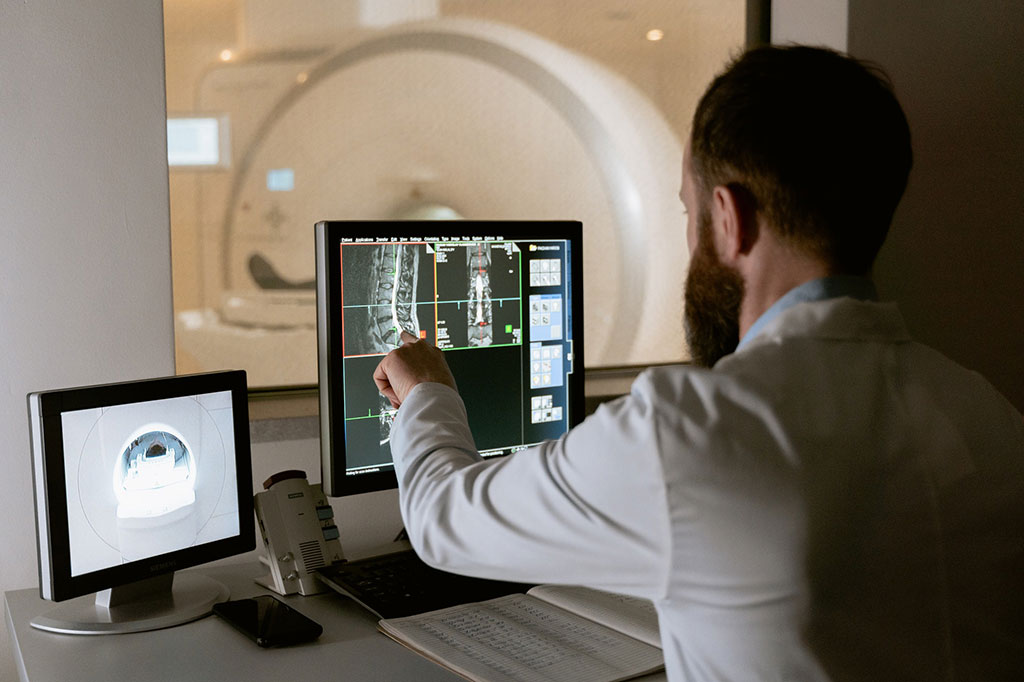
New AI-Based Image Analysis Platform Enables Physicians to Analyze CT and MRI Scans in Half the Time of Traditional Methods
|
By MedImaging International staff writers Posted on 21 Jan 2021 |


|
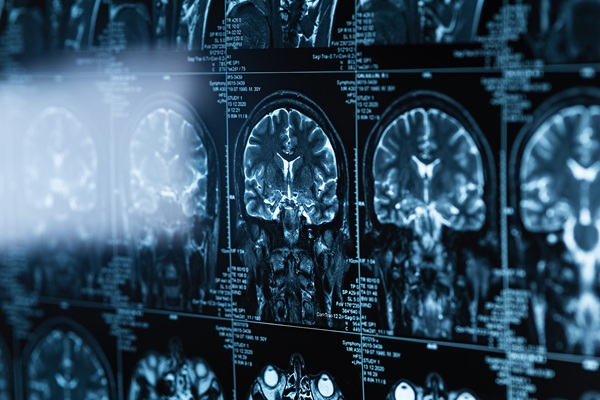
Illustration
The US Food and Drug Administration (FDA) has granted 510(k) clearance for a new image analysis platform that enables physicians to analyze CT and MRI scans in half the time of traditional methods.
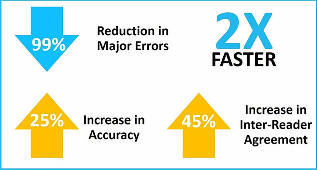
The new image analysis platform named AI Mass has been launched by AI Metrics, LLC (Birmingham, AL, USA) which has successfully implemented augmented intelligence - the concept of using artificial intelligence (AI) to improve human performance. The AI Metrics platform utilizes AI-assisted workflows to improve accuracy and consistency, and dramatically reduce errors.
The first application on AI Mass assists radiologists with image analysis and reporting of advanced cancer over time. During clinical trials for novel therapeutics, a patient’s response to therapy is assessed over time using detailed criteria. Such criteria require highly-controlled tumor measurements and calculations of percent changes in tumor size. In clinical practice, assessments are more subjective and lack standardization due to time constraints placed upon physicians.
“Our goal was to design a simple, intuitive platform in which radiologists could assess medical images from patients with minimal errors and a high level of accuracy, consistency, and efficiency,” said Dr. Andrew Smith, CEO and Founder of AI Metrics. “To do this, we had to break free from outdated methods and create an entirely new image viewer that supports AI and best-practice workflows that generate crystal clear data and reports. We started with advanced cancer and lymphoma, and are now broadening our platform to include early-stage cancers.”
“We solved the efficiency and standardization with a combination of AI and guided workflows that capture the best aspects of clinical trials and bring them into clinical practice,” added Smith. “This advanced technology completely changes the standard of care - for radiologists, oncologists and patients. As a practicing radiologist and researcher, I’ve envisioned for some time being able to use a system like this, and I am excited to see years of design work finally materialize into an incredible product.”
Related Links:
AI Metrics, LLC




The new image analysis platform named AI Mass has been launched by AI Metrics, LLC (Birmingham, AL, USA) which has successfully implemented augmented intelligence - the concept of using artificial intelligence (AI) to improve human performance. The AI Metrics platform utilizes AI-assisted workflows to improve accuracy and consistency, and dramatically reduce errors.
The first application on AI Mass assists radiologists with image analysis and reporting of advanced cancer over time. During clinical trials for novel therapeutics, a patient’s response to therapy is assessed over time using detailed criteria. Such criteria require highly-controlled tumor measurements and calculations of percent changes in tumor size. In clinical practice, assessments are more subjective and lack standardization due to time constraints placed upon physicians.
“Our goal was to design a simple, intuitive platform in which radiologists could assess medical images from patients with minimal errors and a high level of accuracy, consistency, and efficiency,” said Dr. Andrew Smith, CEO and Founder of AI Metrics. “To do this, we had to break free from outdated methods and create an entirely new image viewer that supports AI and best-practice workflows that generate crystal clear data and reports. We started with advanced cancer and lymphoma, and are now broadening our platform to include early-stage cancers.”
“We solved the efficiency and standardization with a combination of AI and guided workflows that capture the best aspects of clinical trials and bring them into clinical practice,” added Smith. “This advanced technology completely changes the standard of care - for radiologists, oncologists and patients. As a practicing radiologist and researcher, I’ve envisioned for some time being able to use a system like this, and I am excited to see years of design work finally materialize into an incredible product.”
Related Links:
AI Metrics, LLC

Post-Processing Imaging System
DynaCAD Prostate
X-ray Diagnostic System
FDX Visionary-A
Biopsy Software
Affirm® Contrast
Ultrasound Table
Women’s Ultrasound EA Table
Latest Industry News News
- GE HealthCare Highlights AI-Supported Radiation Therapy Tools at ESTRO 2026
- Nuclear Medicine Set for Continued Growth Driven by Demand for Precision Diagnostics
- GE HealthCare and NVIDIA Collaboration to Reimagine Diagnostic Imaging
- Patient-Specific 3D-Printed Phantoms Transform CT Imaging
- Siemens and Sectra Collaborate on Enhancing Radiology Workflows
- Bracco Diagnostics and ColoWatch Partner to Expand Availability CRC Screening Tests Using Virtual Colonoscopy
- Mindray Partners with TeleRay to Streamline Ultrasound Delivery
- Philips and Medtronic Partner on Stroke Care
- Siemens and Medtronic Enter into Global Partnership for Advancing Spine Care Imaging Technologies
- RSNA 2024 Technical Exhibits to Showcase Latest Advances in Radiology
- Bracco Collaborates with Arrayus on Microbubble-Assisted Focused Ultrasound Therapy for Pancreatic Cancer
- Innovative Collaboration to Enhance Ischemic Stroke Detection and Elevate Standards in Diagnostic Imaging
- RSNA 2024 Registration Opens
- Microsoft collaborates with Leading Academic Medical Systems to Advance AI in Medical Imaging
- GE HealthCare Acquires Intelligent Ultrasound Group’s Clinical Artificial Intelligence Business
- Bayer and Rad AI Collaborate on Expanding Use of Cutting Edge AI Radiology Operational Solutions
Channels
Radiography
view channel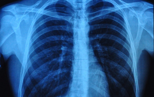
Simple Chest X-Ray Measure Predicts Survival After Lung Cancer Surgery
Obstructive ventilatory disorder, marked by airflow limitation that reduces breathing efficiency, increases postoperative risk in patients with lung cancer. Although surgery offers the best chance of cure,... Read more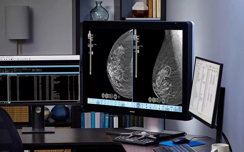
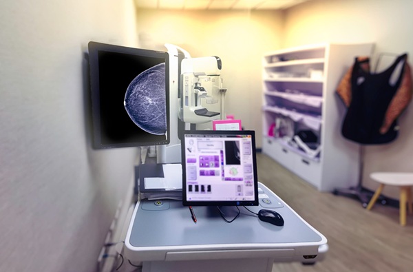
AI Detection Tool Improves Identification of Lobular Breast Cancer
Breast cancer screening seeks early detection, yet some subtypes remain difficult to visualize on mammography, risking delayed diagnosis. On average, 1 in 20 women worldwide will develop breast cancer,... Read more
MRI
view channel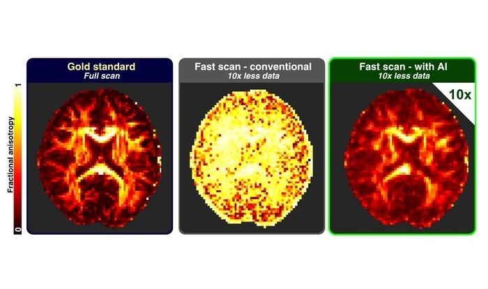
AI Approach Could Shorten Advanced Brain MRI Scans by Up to 90%
Long acquisition times for advanced brain magnetic resonance imaging (MRI) can limit access, extend waiting lists, and disrupt clinical workflows. Reducing data requirements without sacrificing image fidelity... Read more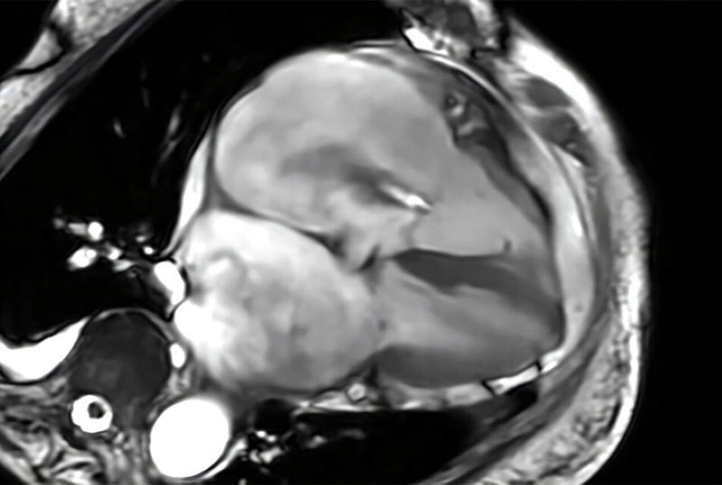
Cardiac MRI Measure Improves Risk Prediction in Tricuspid Regurgitation
Tricuspid regurgitation, in which blood flows back from the right ventricle into the right atrium, can lead to progressive right-sided heart failure. Clinicians need reliable ways to gauge severity and... Read more
Ultrasound
view channel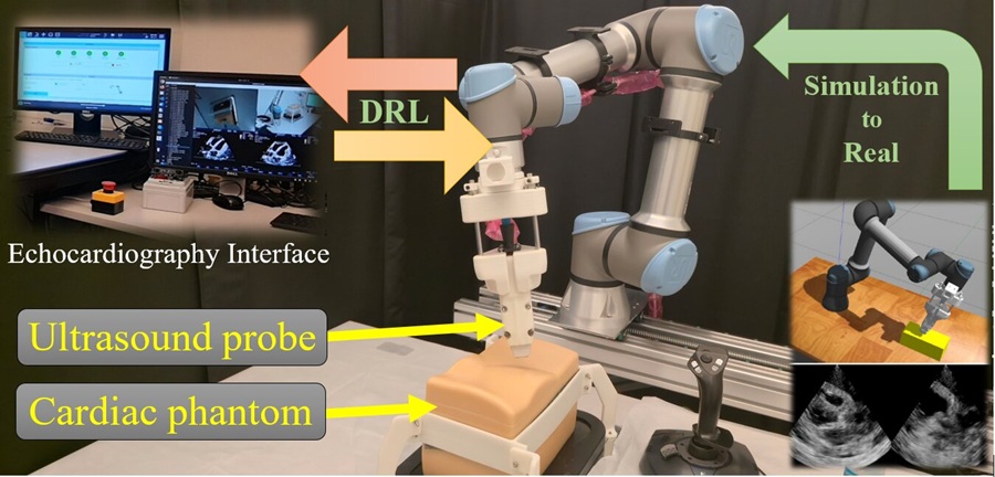
AI Robotic Ultrasound System Automates Echocardiography and Improves Consistency
Echocardiography, an ultrasound examination of the heart, is central to diagnosing and managing cardiovascular disease. Many services struggle with limited availability of skilled sonographers, variable... Read more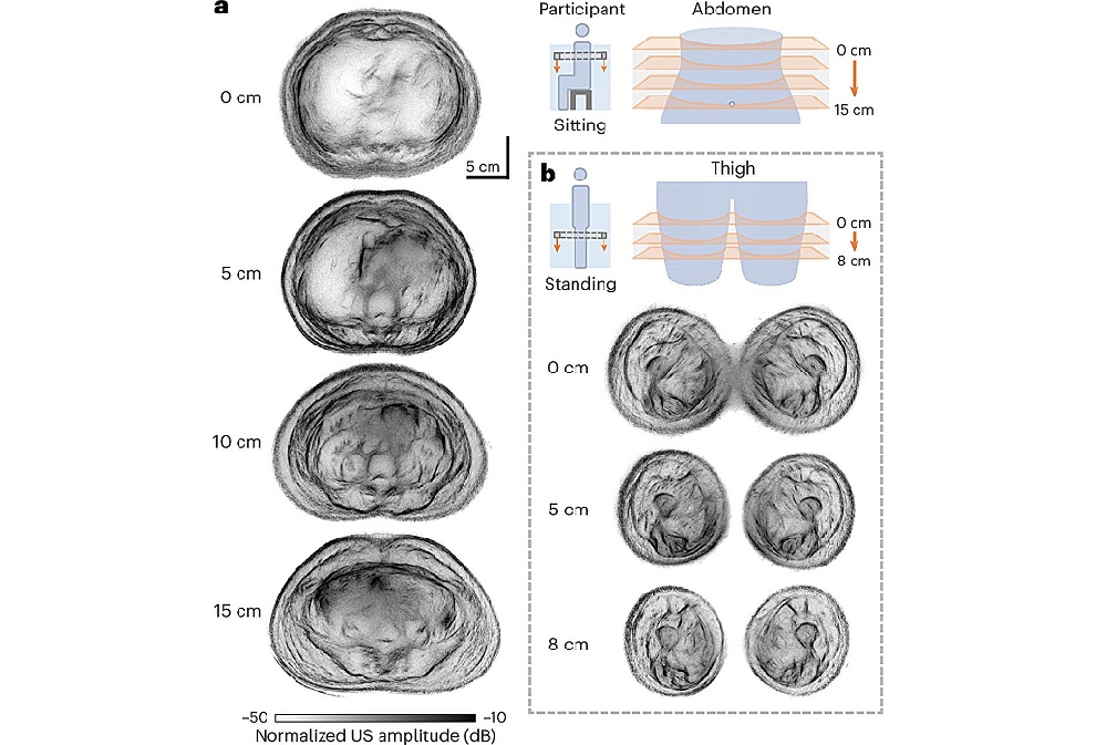
Whole Cross-Section Ultrasound System Enables Operator-Independent Imaging
Conventional ultrasound is central to bedside imaging but is limited by a narrow field of view and operator variability. Comprehensive cross-sectional assessment typically requires computed tomography... Read more
Nuclear Medicine
view channel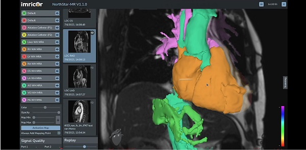
MR-Guided Cardiac Mapping System Enables Radiation-Free Procedures
Cardiac electrophysiology procedures are typically guided by X-ray fluoroscopy, which limits soft-tissue visualization and exposes patients and clinical staff to ionizing radiation. Real-time mapping that... Read more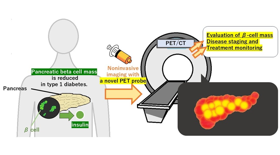
PET Tracer Enables Noninvasive Measurement of Beta Cell Mass
Type 1 diabetes is an autoimmune disease in which the immune system destroys insulin-producing pancreatic beta cells. Loss of these cells destabilizes glucose control and drives complications.... Read more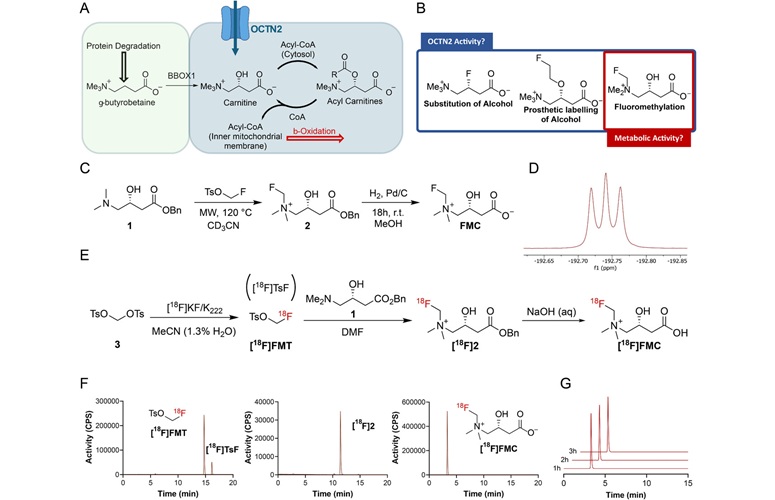
New Imaging Tool Sheds Light on Tumor Fat Metabolism
Rapidly growing tumors reprogram metabolism to meet high energy demands. While many cancers preferentially consume glucose, lipid utilization by malignant cells is difficult to measure in living subjects.... Read more
Radiopharmaceutical Molecule Marker to Improve Choice of Bladder Cancer Therapies
Targeted cancer therapies only work when tumor cells express the specific molecular structures they are designed to attack. In urothelial carcinoma, a common form of bladder cancer, the cell surface protein... Read moreGeneral/Advanced Imaging
view channel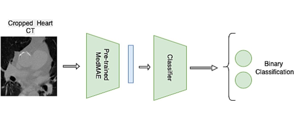
Multimodal AI Tool Combines CT and Health Records to Predict Heart Risk
Cardiovascular disease is a leading cause of death and an underrecognized risk for people treated for breast cancer. Cardiac complications can affect survival and quality of life. Clinicians need tools... Read more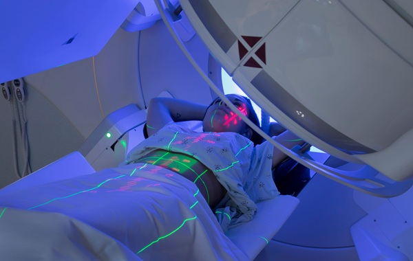
AI Tool Automates Radiotherapy Planning for Cervical and Prostate Cancer
Cervical cancer causes most of its global mortality in low- and middle-income countries, where radiotherapy capacity and specialist staff are limited. Treatment planning is labor-intensive and can delay... Read more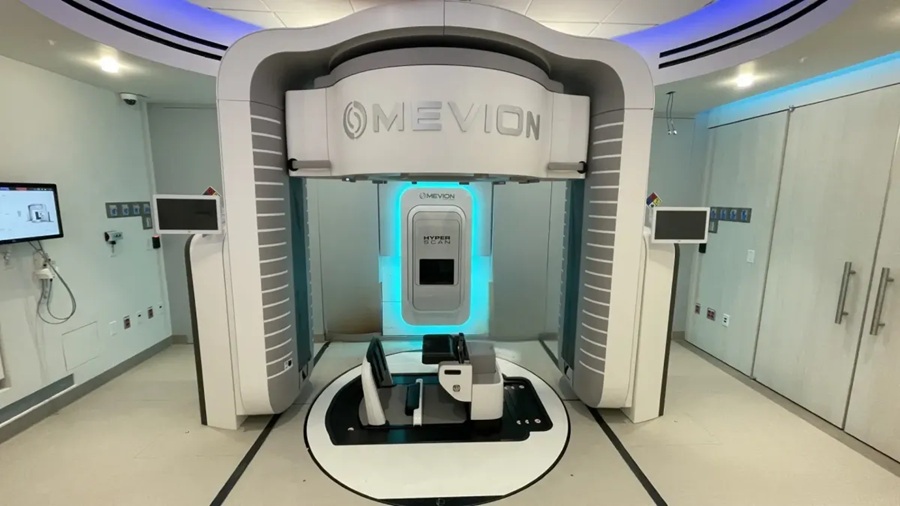
Imaging IT
view channel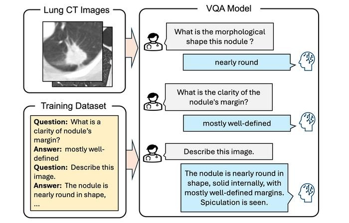
Interactive AI Tool Supports Explainable Lung Nodule Assessment
Lung cancer is a leading cause of cancer mortality, and timely characterization of pulmonary nodules on chest computed tomography (CT) is essential for directing care. Interpreting nodule morphology demands... Read more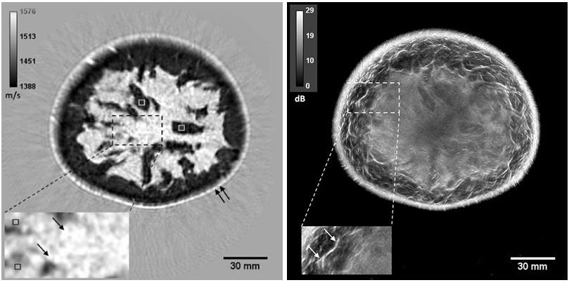
Breast Imaging Software Enhances Visualization and Tissue Characterization in Challenging Cases
Breast imaging can be particularly challenging in cases involving small breasts or implants, where image reconstruction and tissue characterization may be limited. Clinicians also need reproducible analysis... Read more
New Google Cloud Medical Imaging Suite Makes Imaging Healthcare Data More Accessible
Medical imaging is a critical tool used to diagnose patients, and there are billions of medical images scanned globally each year. Imaging data accounts for about 90% of all healthcare data1 and, until... Read more