Synergistic Ultrasound Platform Advances Breast Imaging
|
By MedImaging International staff writers Posted on 18 Jan 2021 |
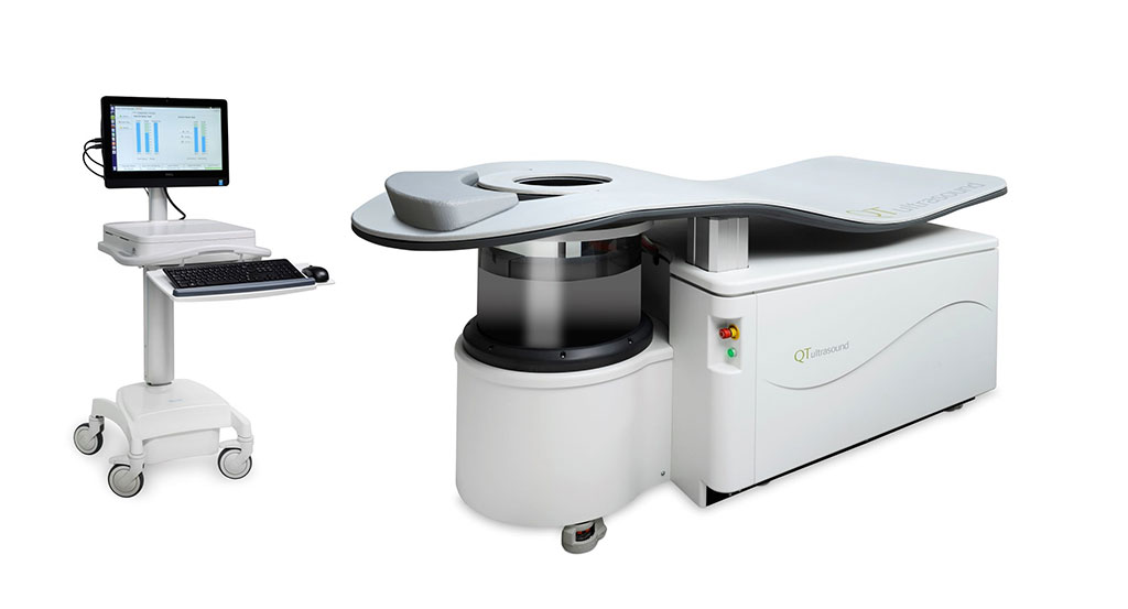
Image: The QTscan and QTviewer (Photo courtesy of QT Imaging)
A new quantitative transmission ultrasound imaging system provides reflection-mode and transmission-mode images of a patient's breasts.
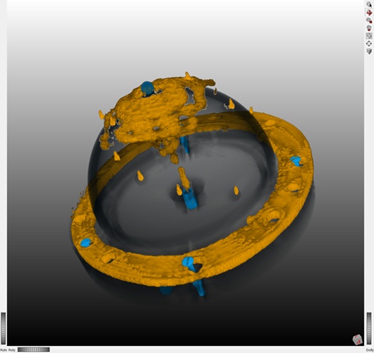
The QT Imaging (Novato, CA, USA) QTscan is a non-invasive breast imaging tool that uses a transmitter/receiver array pair for complementary transmission and reflection ultrasound in order to generate highly accurate three dimensional (3D) volumetric ultrasound images. QTscan consists of a patient support table and a scan tank filled with warm water that holds the ultrasound transducer arrays, and an additional transmitter and receiver array pair to collect ultrasound energy for speed of sound (SOS) values.
The transducer array includes three reflectors that transmit pulsed ultrasound plane waves into targeted tissues, with the water bath in the scan tank as a coupling medium. During scanning, the patient lies prone face down on the support table with the breast suspended in the scan bath, with the nipple as a point of reference. The transducer arrays rotate about a vertical axis to circle the breast in the coronal plane. The array then translates vertically and the scanning process is repeated until the entire breast is scanned.
The QTscan then outputs the B-scan images to be constructively combined into tomographic, SOS, and reflection ultrasound images, which can be viewed on the proprietary QTviewer in coronal, axial, and sagittal sections. SOS images may be queried by probe and region of interest (ROI) tools provided in the viewer console that provide SOS values in meters/sec to aid in the diagnostic evaluation of the breast.
“The QTscan allows for painless breast imaging, as no compression is required, with no need for x-ray radiation (such as in mammography) and no need for administration of contrast agents, as required for the majority of breast MRI scans,” said the company. “It is intended to be used as an initial evaluation method for asymptomatic women identified with above-average risk for developing breast cancer, based on genetic testing or other established criteria. It is not intended to be used as a replacement for standard screening mammography.”
Related Links:
QT Imaging
Latest Ultrasound News
- Non-Invasive Ultrasound-Based Tool Accurately Detects Infant Meningitis
- Breakthrough Deep Learning Model Enhances Handheld 3D Medical Imaging
- Pain-Free Breast Imaging System Performs One Minute Cancer Scan
- Wireless Chronic Pain Management Device to Reduce Need for Painkillers and Surgery
- New Medical Ultrasound Imaging Technique Enables ICU Bedside Monitoring
- New Incision-Free Technique Halts Growth of Debilitating Brain Lesions
- AI-Powered Lung Ultrasound Outperforms Human Experts in Tuberculosis Diagnosis
- AI Identifies Heart Valve Disease from Common Imaging Test
- Novel Imaging Method Enables Early Diagnosis and Treatment Monitoring of Type 2 Diabetes
- Ultrasound-Based Microscopy Technique to Help Diagnose Small Vessel Diseases
- Smart Ultrasound-Activated Immune Cells Destroy Cancer Cells for Extended Periods
- Tiny Magnetic Robot Takes 3D Scans from Deep Within Body
- High Resolution Ultrasound Speeds Up Prostate Cancer Diagnosis
- World's First Wireless, Handheld, Whole-Body Ultrasound with Single PZT Transducer Makes Imaging More Accessible
- Artificial Intelligence Detects Undiagnosed Liver Disease from Echocardiograms
- Ultrasound Imaging Non-Invasively Tracks Tumor Response to Radiation and Immunotherapy
Channels
Radiography
view channel
AI Hybrid Strategy Improves Mammogram Interpretation
Breast cancer screening programs rely heavily on radiologists interpreting mammograms, a process that is time-intensive and subject to errors. While artificial intelligence (AI) models have shown strong... Read more
AI Technology Predicts Personalized Five-Year Risk of Developing Breast Cancer
Breast cancer remains one of the most common cancers among women, with about one in eight receiving a diagnosis in their lifetime. Despite widespread use of mammography, about 34% of patients in the U.... Read moreMRI
view channel
AI-Assisted Model Enhances MRI Heart Scans
A cardiac MRI can reveal critical information about the heart’s function and any abnormalities, but traditional scans take 30 to 90 minutes and often suffer from poor image quality due to patient movement.... Read more
AI Model Outperforms Doctors at Identifying Patients Most At-Risk of Cardiac Arrest
Hypertrophic cardiomyopathy is one of the most common inherited heart conditions and a leading cause of sudden cardiac death in young individuals and athletes. While many patients live normal lives, some... Read moreNuclear Medicine
view channel
New Camera Sees Inside Human Body for Enhanced Scanning and Diagnosis
Nuclear medicine scans like single-photon emission computed tomography (SPECT) allow doctors to observe heart function, track blood flow, and detect hidden diseases. However, current detectors are either... Read more
Novel Bacteria-Specific PET Imaging Approach Detects Hard-To-Diagnose Lung Infections
Mycobacteroides abscessus is a rapidly growing mycobacteria that primarily affects immunocompromised patients and those with underlying lung diseases, such as cystic fibrosis or chronic obstructive pulmonary... Read moreGeneral/Advanced Imaging
view channel
Extending CT Imaging Detects Hidden Blood Clots in Stroke Patients
Strokes caused by blood clots or other mechanisms that obstruct blood flow in the brain account for about 85% of all strokes. Determining where a clot originates is crucial, since it guides safe and effective... Read more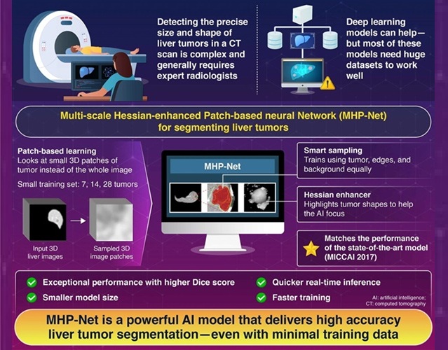
Groundbreaking AI Model Accurately Segments Liver Tumors from CT Scans
Liver cancer is the sixth most common cancer worldwide and a leading cause of cancer-related deaths. Accurate segmentation of liver tumors is critical for diagnosis and therapy, but manual methods by radiologists... Read more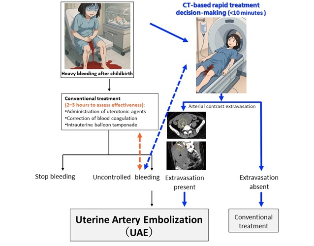
New CT-Based Indicator Helps Predict Life-Threatening Postpartum Bleeding Cases
Postpartum hemorrhage (PPH) is a leading cause of maternal death worldwide. While most cases can be controlled with medications and basic interventions, some become life-threatening and require invasive treatments.... Read moreImaging IT
view channel
New Google Cloud Medical Imaging Suite Makes Imaging Healthcare Data More Accessible
Medical imaging is a critical tool used to diagnose patients, and there are billions of medical images scanned globally each year. Imaging data accounts for about 90% of all healthcare data1 and, until... Read more
Global AI in Medical Diagnostics Market to Be Driven by Demand for Image Recognition in Radiology
The global artificial intelligence (AI) in medical diagnostics market is expanding with early disease detection being one of its key applications and image recognition becoming a compelling consumer proposition... Read moreIndustry News
view channel
GE HealthCare and NVIDIA Collaboration to Reimagine Diagnostic Imaging
GE HealthCare (Chicago, IL, USA) has entered into a collaboration with NVIDIA (Santa Clara, CA, USA), expanding the existing relationship between the two companies to focus on pioneering innovation in... Read more
Patient-Specific 3D-Printed Phantoms Transform CT Imaging
New research has highlighted how anatomically precise, patient-specific 3D-printed phantoms are proving to be scalable, cost-effective, and efficient tools in the development of new CT scan algorithms... Read more
Siemens and Sectra Collaborate on Enhancing Radiology Workflows
Siemens Healthineers (Forchheim, Germany) and Sectra (Linköping, Sweden) have entered into a collaboration aimed at enhancing radiologists' diagnostic capabilities and, in turn, improving patient care... Read more


 Guided Devices.jpg)


