MRI Shows Brain Anatomy Differences Between Autistic and Typically Developing Individuals to Be Mostly Indistinguishable
|
By MedImaging International staff writers Posted on 24 Nov 2014 |

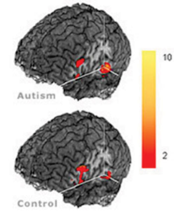
Image: Differences in the brains of autistic and control subjects using MRI (Photo courtesy of Center of Cognitive Brain Imaging, Carnegie Mellon University).

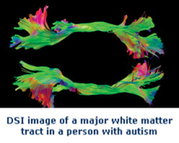
Image: MR diffusion spectrum imaging (DSI) used to examine the structural integrity of white matter in people with autism and typical participants. These imaging techniques visualize axonal tracts by measuring the diffusion of water along white matter fibers (Photo courtesy of Center of Cognitive Brain Imaging, Carnegie Mellon University).
In the largest magnetic resonance imaging (MRI) study of patients with autism to date, Israeli and American researchers have shown that the brain anatomy in MRI scans of autistic individuals older than age six is mostly indistinguishable from that of typically developing individuals, and therefore, of little clinical or scientific value.
The researchers from Ben-Gurion University of the Negev (BGU; Be’er Sheva, Israel) and Carnegie Mellon University (Pittsburgh, PA, USA) published their findings online October 14, 2014, in the Oxford journal Cerebral Cortex. “Our findings offer definitive answers regarding several scientific controversies about brain anatomy, which have occupied autism research for the past 10 to 15 years,” said Dr. Ilan Dinstein of BGU’s departments of psychology and brain and cognitive sciences. “Previous hypotheses suggesting that autism is associated with larger intracranial gray matter, white matter, and amygdala volumes, or smaller cerebellar, corpus callosum and hippocampus volumes were mostly refuted by this new study.”
The researchers used data from the Autism Brain Imaging Data Exchange (ABIDE), which provides an unprecedented prospect to conduct large-scale comparisons of anatomic MRI scans across autism and control groups and resolve many outstanding questions. This recently released database is a worldwide collection of MRI scans from over 1,000 individuals (half with autism and half controls) ages 6 to 35 years old.
“In the study we performed very detailed anatomical examinations of the scans, which included dividing each brain into over 180 regions of interest and assessing multiple anatomical measures such as the volume, surface area and thickness of each region,” Dr. Dinstein stated.
The researchers then examined how the autism and control groups differed with respect to each region and also with respect to groups of regions using more complex analyses. “The most striking finding here was that anatomical differences within both the control group and the autistic group was immense and greatly overshadowed minute differences between the two groups,” Dr. Dinstein noted. “For example, individuals in the control group differ by 80% to 90% in their brain volumes, while differences in brain volume across autism and control groups differed by 2% to 3% percent at most. This led us to the conclusion that anatomical measures of brain volume or surface areas do not offer much information regarding the underlying mechanism or pathology of autistic spectrum disorder [ASD],” he stated. “These sobering results suggest that autism is not a disorder that is associated with specific anatomical pathology and as a result, anatomical measures alone are likely to be of low scientific and clinical significance for identifying children, adolescents and adults with ASD, or for elucidating their neuropathology.”
Dr. Dinstein believes that more complicated explanations involving combinations of measures in more homogeneous subgroups are in all probability to be the answer. “Expecting to find a single answer for the entire ASD population is naive. We need to move on to thinking about how to split up this very heterogeneous group of disorders into more meaningful biologically-relevant subgroups,” he stated.
This conclusion stands in great contrast to numerous reports of significant anatomical differences described by smaller studies, which have typically included comparisons of 40 to 50 individuals. “The problem with small samples, large within-group heterogeneity, and a scientific bias to report only positive findings, is that small samples are likely to yield significant differences across autism and control groups in a few of the 180 brain regions,” Dr. Dinstein explained.
“In such a situation one would expect that each study would find significant differences in different brain areas and that findings will be very inconsistent across studies," he says. "This is exactly what you see when you examine the autism anatomy literature from the last decade or so. Our study simply explains why this has been happening and puts an end to several ensuing debates.”
Related Links:
Ben-Gurion University of the Negev
Center of Cognitive Brain Imaging, Carnegie Mellon University
Carnegie Mellon University
Latest MRI News
- MRI Scans Reveal Signature Patterns of Brain Activity to Predict Recovery from TBI
- Novel Imaging Approach to Improve Treatment for Spinal Cord Injuries
- AI-Assisted Model Enhances MRI Heart Scans
- AI Model Outperforms Doctors at Identifying Patients Most At-Risk of Cardiac Arrest
- New MRI Technique Reveals Hidden Heart Issues
- Shorter MRI Exam Effectively Detects Cancer in Dense Breasts
- MRI to Replace Painful Spinal Tap for Faster MS Diagnosis
- MRI Scans Can Identify Cardiovascular Disease Ten Years in Advance
- Simple Brain Scan Diagnoses Parkinson's Disease Years Before It Becomes Untreatable
- Cutting-Edge MRI Technology to Revolutionize Diagnosis of Common Heart Problem
- New MRI Technique Reveals True Heart Age to Prevent Attacks and Strokes
- AI Tool Predicts Relapse of Pediatric Brain Cancer from Brain MRI Scans
- AI Tool Tracks Effectiveness of Multiple Sclerosis Treatments Using Brain MRI Scans
- Ultra-Powerful MRI Scans Enable Life-Changing Surgery in Treatment-Resistant Epileptic Patients
- AI-Powered MRI Technology Improves Parkinson’s Diagnoses
- Biparametric MRI Combined with AI Enhances Detection of Clinically Significant Prostate Cancer
Channels
Radiography
view channel
Routine Mammograms Could Predict Future Cardiovascular Disease in Women
Mammograms are widely used to screen for breast cancer, but they may also contain overlooked clues about cardiovascular health. Calcium deposits in the arteries of the breast signal stiffening blood vessels,... Read more
AI Detects Early Signs of Aging from Chest X-Rays
Chronological age does not always reflect how fast the body is truly aging, and current biological age tests often rely on DNA-based markers that may miss early organ-level decline. Detecting subtle, age-related... Read moreUltrasound
view channel
Wearable Ultrasound Imaging System to Enable Real-Time Disease Monitoring
Chronic conditions such as hypertension and heart failure require close monitoring, yet today’s ultrasound imaging is largely confined to hospitals and short, episodic scans. This reactive model limits... Read more
Ultrasound Technique Visualizes Deep Blood Vessels in 3D Without Contrast Agents
Producing clear 3D images of deep blood vessels has long been difficult without relying on contrast agents, CT scans, or MRI. Standard ultrasound typically provides only 2D cross-sections, limiting clinicians’... Read moreNuclear Medicine
view channel
PET Imaging of Inflammation Predicts Recovery and Guides Therapy After Heart Attack
Acute myocardial infarction can trigger lasting heart damage, yet clinicians still lack reliable tools to identify which patients will regain function and which may develop heart failure.... Read more
Radiotheranostic Approach Detects, Kills and Reprograms Aggressive Cancers
Aggressive cancers such as osteosarcoma and glioblastoma often resist standard therapies, thrive in hostile tumor environments, and recur despite surgery, radiation, or chemotherapy. These tumors also... Read more
New Imaging Solution Improves Survival for Patients with Recurring Prostate Cancer
Detecting recurrent prostate cancer remains one of the most difficult challenges in oncology, as standard imaging methods such as bone scans and CT scans often fail to accurately locate small or early-stage tumors.... Read moreGeneral/Advanced Imaging
view channel
AI-Based Tool Accelerates Detection of Kidney Cancer
Diagnosing kidney cancer depends on computed tomography scans, often using contrast agents to reveal abnormalities in kidney structure. Tumors are not always searched for deliberately, as many scans are... Read more
New Algorithm Dramatically Speeds Up Stroke Detection Scans
When patients arrive at emergency rooms with stroke symptoms, clinicians must rapidly determine whether the cause is a blood clot or a brain bleed, as treatment decisions depend on this distinction.... Read moreImaging IT
view channel
New Google Cloud Medical Imaging Suite Makes Imaging Healthcare Data More Accessible
Medical imaging is a critical tool used to diagnose patients, and there are billions of medical images scanned globally each year. Imaging data accounts for about 90% of all healthcare data1 and, until... Read more
Global AI in Medical Diagnostics Market to Be Driven by Demand for Image Recognition in Radiology
The global artificial intelligence (AI) in medical diagnostics market is expanding with early disease detection being one of its key applications and image recognition becoming a compelling consumer proposition... Read moreIndustry News
view channel
GE HealthCare and NVIDIA Collaboration to Reimagine Diagnostic Imaging
GE HealthCare (Chicago, IL, USA) has entered into a collaboration with NVIDIA (Santa Clara, CA, USA), expanding the existing relationship between the two companies to focus on pioneering innovation in... Read more
Patient-Specific 3D-Printed Phantoms Transform CT Imaging
New research has highlighted how anatomically precise, patient-specific 3D-printed phantoms are proving to be scalable, cost-effective, and efficient tools in the development of new CT scan algorithms... Read more
Siemens and Sectra Collaborate on Enhancing Radiology Workflows
Siemens Healthineers (Forchheim, Germany) and Sectra (Linköping, Sweden) have entered into a collaboration aimed at enhancing radiologists' diagnostic capabilities and, in turn, improving patient care... Read more


