Imaging Shows Unique Brain Region Tied to Higher Cognitive Abilities
|
By MedImaging International staff writers Posted on 18 Feb 2014 |

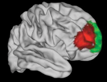
Image: An area (red) of the brain that seems to be unique to humans (Photo courtesy of the University of Oxford).
British researchers have pinpointed an area of the human brain that appears unlike anything in the brains of some of human’s closest relatives. The brain area identified is known to be closely involved in some of the most advanced planning and decision-making processes that people think of as being expressly human.
“We tend to think that being able to plan into the future, be flexible in our approach and learn from others are things that are particularly impressive about humans. We’ve identified an area of the brain that appears to be uniquely human and is likely to have something to do with these cognitive powers,” said senior researcher Prof. Matthew Rushworth , from the department of experimental psychology at Oxford University’s (UK).
Magnetic resonance imaging (MRI) imaging of 25 adult volunteers was used to identify key components in the ventrolateral frontal cortex area of the human brain, and map how these components were connected up with other brain areas. The findings were then compared to equivalent MRI data from 25 macaque monkeys.
Involved in many of the highest facets of cognition and language, the ventrolateral frontal cortex region of the brain is only present in humans and other primates. Some areas are implicated in psychiatric conditions such as drug addiction, attention deficit hyperactivity disorder (ADHD), or compulsive behavior disorders. Language is affected when other areas are damaged after stroke or neurodegenerative disease. A better determination of the neural connections and networks involved should help the understanding of alterations in the brain that are linked with these disorders.
The Oxford University researchers reported their findings February 5, 2014, in the journal Neuron. Prof. Rushworth clarified, “The brain is a mosaic of interlinked areas. We wanted to look at this very important region of the frontal part of the brain and see how many tiles there are and where they are placed. We also looked at the connections of each tile—how they are wired up to the rest of the brain—as it is these connections that determine the information that can reach that component part and the influence that part can have on other brain regions.”
The researchers, utilizing the MRI data, were able to divide the human ventrolateral frontal cortex into 12 areas that were consistent across all the individuals. “Each of these 12 areas has its own pattern of connections with the rest of the brain, a sort of ‘neural fingerprint,’ telling us it is doing something unique,” said Prof. Rushworth.
The researchers were then able to compare the 12 areas in the human brain region with the organization of the monkey prefrontal cortex. Overall, they were very similar with 11 of the 12 areas being found in both species and being connected up to other brain areas in very similar ways. However, one area of the human ventrolateral frontal cortex had no corresponding area in the macaque—a region called the lateral frontal pole prefrontal cortex. “We have established an area in human frontal cortex which does not seem to have an equivalent in the monkey at all,” stated first author Oxford’s Franz-Xaver Neubert. “This area has been identified with strategic planning and decision making as well as multitasking.”
Moreover, the researchers revealed that the auditory areas of the brain were very well connected with the human prefrontal cortex, but much less so in the macaque. The researchers suggest this may be essential for our ability to understand and generate speech.
Related Links:
Oxford University
Latest MRI News
- MRI Scan Breakthrough to Help Avoid Risky Invasive Tests for Heart Patients
- MRI Scans Reveal Signature Patterns of Brain Activity to Predict Recovery from TBI
- Novel Imaging Approach to Improve Treatment for Spinal Cord Injuries
- AI-Assisted Model Enhances MRI Heart Scans
- AI Model Outperforms Doctors at Identifying Patients Most At-Risk of Cardiac Arrest
- New MRI Technique Reveals Hidden Heart Issues
- Shorter MRI Exam Effectively Detects Cancer in Dense Breasts
- MRI to Replace Painful Spinal Tap for Faster MS Diagnosis
- MRI Scans Can Identify Cardiovascular Disease Ten Years in Advance
- Simple Brain Scan Diagnoses Parkinson's Disease Years Before It Becomes Untreatable
- Cutting-Edge MRI Technology to Revolutionize Diagnosis of Common Heart Problem
- New MRI Technique Reveals True Heart Age to Prevent Attacks and Strokes
- AI Tool Predicts Relapse of Pediatric Brain Cancer from Brain MRI Scans
- AI Tool Tracks Effectiveness of Multiple Sclerosis Treatments Using Brain MRI Scans
- Ultra-Powerful MRI Scans Enable Life-Changing Surgery in Treatment-Resistant Epileptic Patients
- AI-Powered MRI Technology Improves Parkinson’s Diagnoses
Channels
Radiography
view channel
Routine Mammograms Could Predict Future Cardiovascular Disease in Women
Mammograms are widely used to screen for breast cancer, but they may also contain overlooked clues about cardiovascular health. Calcium deposits in the arteries of the breast signal stiffening blood vessels,... Read more
AI Detects Early Signs of Aging from Chest X-Rays
Chronological age does not always reflect how fast the body is truly aging, and current biological age tests often rely on DNA-based markers that may miss early organ-level decline. Detecting subtle, age-related... Read moreUltrasound
view channel
Portable Ultrasound Sensor to Enable Earlier Breast Cancer Detection
Breast cancer screening relies heavily on annual mammograms, but aggressive tumors can develop between scans, accounting for up to 30 percent of cases. These interval cancers are often diagnosed later,... Read more
Portable Imaging Scanner to Diagnose Lymphatic Disease in Real Time
Lymphatic disorders affect hundreds of millions of people worldwide and are linked to conditions ranging from limb swelling and organ dysfunction to birth defects and cancer-related complications.... Read more
Imaging Technique Generates Simultaneous 3D Color Images of Soft-Tissue Structure and Vasculature
Medical imaging tools often force clinicians to choose between speed, structural detail, and functional insight. Ultrasound is fast and affordable but typically limited to two-dimensional anatomy, while... Read moreNuclear Medicine
view channel
Radiopharmaceutical Molecule Marker to Improve Choice of Bladder Cancer Therapies
Targeted cancer therapies only work when tumor cells express the specific molecular structures they are designed to attack. In urothelial carcinoma, a common form of bladder cancer, the cell surface protein... Read more
Cancer “Flashlight” Shows Who Can Benefit from Targeted Treatments
Targeted cancer therapies can be highly effective, but only when a patient’s tumor expresses the specific protein the treatment is designed to attack. Determining this usually requires biopsies or advanced... Read moreGeneral/Advanced Imaging
view channel
AI Tool Offers Prognosis for Patients with Head and Neck Cancer
Oropharyngeal cancer is a form of head and neck cancer that can spread through lymph nodes, significantly affecting survival and treatment decisions. Current therapies often involve combinations of surgery,... Read more
New 3D Imaging System Addresses MRI, CT and Ultrasound Limitations
Medical imaging is central to diagnosing and managing injuries, cancer, infections, and chronic diseases, yet existing tools each come with trade-offs. Ultrasound, X-ray, CT, and MRI can be costly, time-consuming,... Read moreImaging IT
view channel
New Google Cloud Medical Imaging Suite Makes Imaging Healthcare Data More Accessible
Medical imaging is a critical tool used to diagnose patients, and there are billions of medical images scanned globally each year. Imaging data accounts for about 90% of all healthcare data1 and, until... Read more
Global AI in Medical Diagnostics Market to Be Driven by Demand for Image Recognition in Radiology
The global artificial intelligence (AI) in medical diagnostics market is expanding with early disease detection being one of its key applications and image recognition becoming a compelling consumer proposition... Read moreIndustry News
view channel
Nuclear Medicine Set for Continued Growth Driven by Demand for Precision Diagnostics
Clinical imaging services face rising demand for precise molecular diagnostics and targeted radiopharmaceutical therapy as cancer and chronic disease rates climb. A new market analysis projects rapid expansion... Read more


