Mobile MRI Unit Reveals Effects of Endurance Running
|
By MedImaging International staff writers Posted on 30 Dec 2010 |
Using a mobile magnetic resonance imaging (MRI) unit, researchers followed runners for two months along a 4,500-km course to examine how their bodies responded to the high-stress conditions of an ultra-long-distance race.
The study's findings were presented at the annual meeting of the Radiological Society of North America (RSNA), November 2010 in Chicago, IL, USA. "Due to the exceptional setting of this study, we could acquire huge amounts of unique data regarding how endurance running affects the body's muscle and body fat,” said Uwe Schütz, MD, a specialist in orthopedics and trauma surgery in the department of diagnostic and interventional radiology at the University Hospital of Ulm (Germany). "Much of what we have learned so far can also be applied to the average runner.”
The TransEurope-FootRace 2009 took place during April 19 to June 21, 2009. It started in southern Italy and traversed approximately 4,488 kilometers to the North Cape in Norway. Forty-four of the runners (66%) agreed to participate in the study. Urine and blood samples as well as biometric data were collected daily. The runners were also randomly assigned to other exams, including electrocardiograms, during the course of the study. Twenty-two of the runners in the study underwent a whole-body MRI exam approximately every three or four days during the race, totaling 15 to 17 exams over a period of 64 days. At the close of the race, researchers began to evaluate the data to determine, among other things, stress-induced changes in the legs and feet from running. Whole-body volume, body fat, visceral fat, abdominal subcutaneous adipose tissue (SCAT), and fat and skeletal muscle of the lower extremities were measured. Sophisticated MRI techniques allowed the researchers to quantify muscle tissue, fat, and cartilage changes. According to Dr. Schütz, MRI is the gold standard for the assessment of the musculoskeletal system of the runner.
The MRI scan results demonstrated that runners lost an average of 5.4% body volume during the course of the race, most of which was in the first 2,000 kilometers. They lost 40% of their body fat in the first half of the race and 50% over the duration of the race. Loss of muscle volume in the leg averaged 7%. "One of the surprising things we found is that despite the daily running, the leg muscles of the athletes actually degenerated because of the immense energy consumption,” Dr. Schütz said.
While most people do not run to this extreme, several of the study's other findings still have implications for the marathon runner and even the recreational runner, according to Dr. Schütz. For example, the results showed that some leg injuries are safe to "run through.” If a runner has intermuscular inflammation in the upper or lower legs, it is usually possible to continue running without risk of further tissue damage. Other overuse injuries, such as joint inflammation, carry more risk of progression, but not always with persistent damage. "The rule that ‘if there is pain, you should stop running' is not always correct,” Dr. Schütz said.
Another major finding of the study was that the first tissue affected by running was fat tissue. More significantly, visceral fat loss (mean 70%) occurred much earlier in the running process than previously believed. Visceral fat is the most dangerous fat and is linked to cardiovascular disease. The findings also revealed that the greatest amount of overall fat loss appeared early in the process.
"When you just begin running, the effects of fat reduction are more pronounced than in athletes who have been running their whole life,” Dr. Schütz concluded. "But you should do this sport constantly over the years. If you stop running for a long time, you need to reduce your caloric input or opt for other aerobic exercises to avoid experiencing weight gain.”
Related Links:
University Hospital of Ulm
Latest MRI News
- New MRI Technique Reveals Hidden Heart Issues
- Shorter MRI Exam Effectively Detects Cancer in Dense Breasts
- MRI to Replace Painful Spinal Tap for Faster MS Diagnosis
- MRI Scans Can Identify Cardiovascular Disease Ten Years in Advance
- Simple Brain Scan Diagnoses Parkinson's Disease Years Before It Becomes Untreatable
- Cutting-Edge MRI Technology to Revolutionize Diagnosis of Common Heart Problem
- New MRI Technique Reveals True Heart Age to Prevent Attacks and Strokes
- AI Tool Predicts Relapse of Pediatric Brain Cancer from Brain MRI Scans
- AI Tool Tracks Effectiveness of Multiple Sclerosis Treatments Using Brain MRI Scans
- Ultra-Powerful MRI Scans Enable Life-Changing Surgery in Treatment-Resistant Epileptic Patients
- AI-Powered MRI Technology Improves Parkinson’s Diagnoses
- Biparametric MRI Combined with AI Enhances Detection of Clinically Significant Prostate Cancer
- First-Of-Its-Kind AI-Driven Brain Imaging Platform to Better Guide Stroke Treatment Options
- New Model Improves Comparison of MRIs Taken at Different Institutions
- Groundbreaking New Scanner Sees 'Previously Undetectable' Cancer Spread
- First-Of-Its-Kind Tool Analyzes MRI Scans to Measure Brain Aging
Channels
Radiography
view channel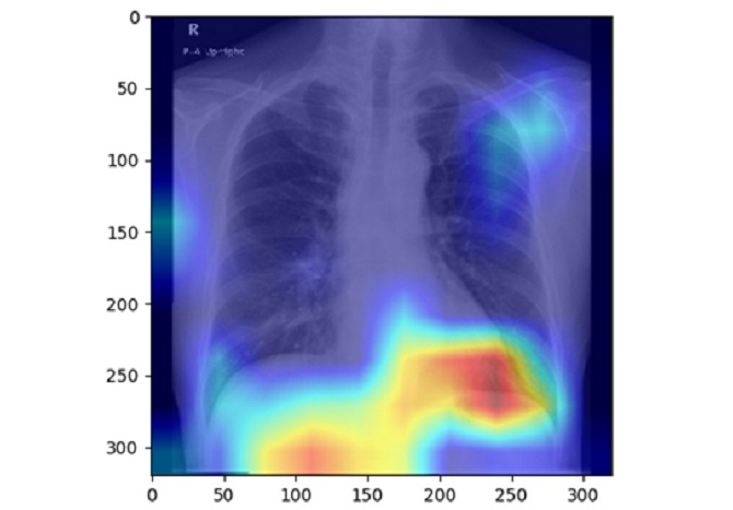
AI Detects Fatty Liver Disease from Chest X-Rays
Fatty liver disease, which results from excess fat accumulation in the liver, is believed to impact approximately one in four individuals globally. If not addressed in time, it can progress to severe conditions... Read more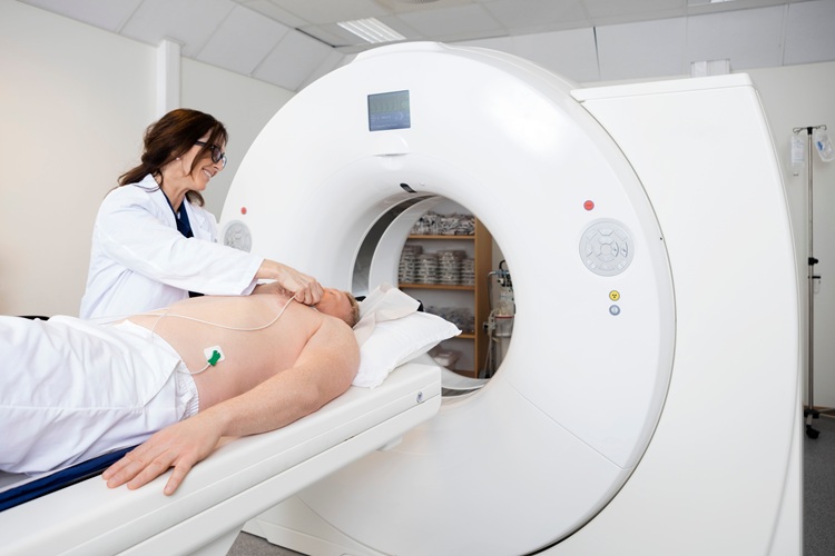
AI Detects Hidden Heart Disease in Existing CT Chest Scans
Coronary artery calcium (CAC) is a major indicator of cardiovascular risk, but its assessment typically requires a specialized “gated” CT scan that synchronizes with the heartbeat. In contrast, most chest... Read moreUltrasound
view channel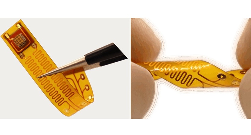
Wireless Chronic Pain Management Device to Reduce Need for Painkillers and Surgery
Chronic pain affects millions of people globally, often leading to long-term disability and dependence on opioid medications, which carry significant risks of side effects and addiction.... Read more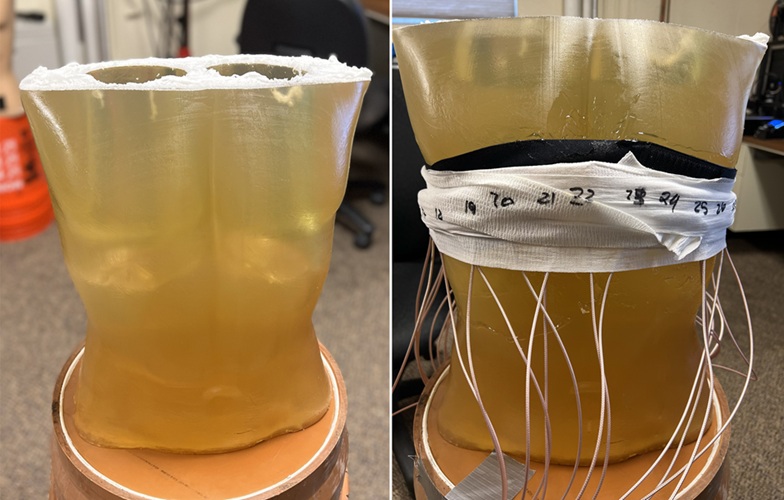
New Medical Ultrasound Imaging Technique Enables ICU Bedside Monitoring
Ultrasound computed tomography (USCT) presents a safer alternative to imaging techniques like X-ray computed tomography (commonly known as CT or “CAT” scans) because it does not produce ionizing radiation.... Read moreNuclear Medicine
view channel
Novel Bacteria-Specific PET Imaging Approach Detects Hard-To-Diagnose Lung Infections
Mycobacteroides abscessus is a rapidly growing mycobacteria that primarily affects immunocompromised patients and those with underlying lung diseases, such as cystic fibrosis or chronic obstructive pulmonary... Read more
New Imaging Approach Could Reduce Need for Biopsies to Monitor Prostate Cancer
Prostate cancer is the second leading cause of cancer-related death among men in the United States. However, the majority of older men diagnosed with prostate cancer have slow-growing, low-risk forms of... Read moreGeneral/Advanced Imaging
view channel
CT Colonography Beats Stool DNA Testing for Colon Cancer Screening
As colorectal cancer remains the second leading cause of cancer-related deaths worldwide, early detection through screening is vital to reduce advanced-stage treatments and associated costs.... Read more
First-Of-Its-Kind Wearable Device Offers Revolutionary Alternative to CT Scans
Currently, patients with conditions such as heart failure, pneumonia, or respiratory distress often require multiple imaging procedures that are intermittent, disruptive, and involve high levels of radiation.... Read more
AI-Based CT Scan Analysis Predicts Early-Stage Kidney Damage Due to Cancer Treatments
Radioligand therapy, a form of targeted nuclear medicine, has recently gained attention for its potential in treating specific types of tumors. However, one of the potential side effects of this therapy... Read moreImaging IT
view channel
New Google Cloud Medical Imaging Suite Makes Imaging Healthcare Data More Accessible
Medical imaging is a critical tool used to diagnose patients, and there are billions of medical images scanned globally each year. Imaging data accounts for about 90% of all healthcare data1 and, until... Read more
Global AI in Medical Diagnostics Market to Be Driven by Demand for Image Recognition in Radiology
The global artificial intelligence (AI) in medical diagnostics market is expanding with early disease detection being one of its key applications and image recognition becoming a compelling consumer proposition... Read moreIndustry News
view channel
GE HealthCare and NVIDIA Collaboration to Reimagine Diagnostic Imaging
GE HealthCare (Chicago, IL, USA) has entered into a collaboration with NVIDIA (Santa Clara, CA, USA), expanding the existing relationship between the two companies to focus on pioneering innovation in... Read more
Patient-Specific 3D-Printed Phantoms Transform CT Imaging
New research has highlighted how anatomically precise, patient-specific 3D-printed phantoms are proving to be scalable, cost-effective, and efficient tools in the development of new CT scan algorithms... Read more
Siemens and Sectra Collaborate on Enhancing Radiology Workflows
Siemens Healthineers (Forchheim, Germany) and Sectra (Linköping, Sweden) have entered into a collaboration aimed at enhancing radiologists' diagnostic capabilities and, in turn, improving patient care... Read more


 Guided Devices.jpg)



.jpeg)


