New Radio-Labeled Molecule Enables Non-Invasive, Real Time Monitoring of Inflammation by PET Imaging
|
By MedImaging International staff writers Posted on 22 Feb 2022 |

A newly developed radio-labeled molecule enables real-time imaging of innate immune activity and offers improved specificity to monitor inflammation across many potential clinical applications.
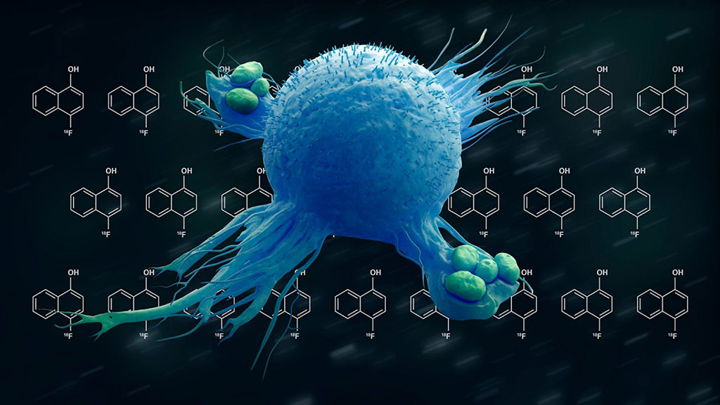
Researchers at The University of Texas MD Anderson Cancer Center (Houston, TX, USA) have developed a new radio-labeled molecule capable of selectively reacting with certain high-energy radicals that are characteristic of innate immune activity, which may allow a non-invasive approach to monitor inflammation in real time by positron emission tomography (PET) imaging. The researchers have taken advantage of new chemistry techniques to synthesize 4-[18F]Fluoro-1-Naphthol ([18F]4FN) as a novel reporter of myeloperoxidase (MPO) activity - a key enzyme active in the innate immune response. The molecule may be able to pinpoint areas of inflammation in a variety of clinical settings, such as inflammatory diseases, infections and immunotherapy-related side effects.
The innate immune response is the body’s first line of defense against invading pathogens. In contrast to the adaptive immune response, innate immunity is nonspecific and acts broadly against infections or foreign agents. Innate immunity is largely driven by myeloid cells, including neutrophils, macrophages and natural killer (NK) cells. Myeloperoxidase is a highly conserved feature of the innate immune response across myeloid cells. This proinflammatory enzyme is activated by hydrogen peroxide to produce a variety of high-energy radicals that are used to eliminate pathogens.
The research team focused on MPO activity to develop a redox-tuned reporter specific to innate immune activity. Using newly developed chemistry techniques, the team was able to synthesize [18F]4FN as a labeled molecule to selectively bind nearby proteins and cells when [18F]4FN has been oxidized by MPO plus hydrogen peroxide, but not hydrogen peroxide alone. The researchers evaluated the potential uses of [18F]4FN as an in vivo PET imaging tool in several laboratory models of inflammation. The molecule was able to successfully highlight inflammation from acute toxic shock, arthritis and contact dermatitis, ailments known to be mediated by activation of innate immunity.
In addition, their results suggest [18F]4FN is a more specific and robust reporter of inflammation than other clinically utilized PET imaging agents, such as fluorodeoxyglucose ([18F]FDG). The research team is in discussions with clinical collaborators to test specific applications of [18F]4FN. An initial study, now under Food and Drug Administration review for Investigational New Drug registration and Institutional Review Board approval, will evaluate [18F]4FN as an early biomarker of immune-related adverse events in patients being treated with immune checkpoint inhibitors.
“There has been a long-standing interest in imaging inflammation and redox in general, but most current approaches generate high levels of background noise from biological processes that generate lower-energy radicals,” said David Piwnica-Worms, M.D., Ph.D., chair of Cancer Systems Imaging. "Our molecule is tuned toward inflammation mediated by high-energy radicals, offering the potential to selectively monitor activation of innate immunity.”
“We need to verify this PET imaging agent in clinical studies, but it certainly has the potential for broad applications that could benefit patients across all kinds of diseases and clinical scenarios,” Piwnica-Worms added. “A tool like this could be used to identify multi-focal hotspots of inflammation, allowing physicians to intervene before disease progression or to follow the resolution of symptoms during therapy.”
Related Links:
The University of Texas MD Anderson Cancer Center
Latest General/Advanced Imaging News
- Radiation Therapy Computed Tomography Solution Boosts Imaging Accuracy
- PET Scans Reveal Hidden Inflammation in Multiple Sclerosis Patients
- Artificial Intelligence Evaluates Cardiovascular Risk from CT Scans
- New AI Method Captures Uncertainty in Medical Images
- CT Coronary Angiography Reduces Need for Invasive Tests to Diagnose Coronary Artery Disease
- Novel Blood Test Could Reduce Need for PET Imaging of Patients with Alzheimer’s
- CT-Based Deep Learning Algorithm Accurately Differentiates Benign From Malignant Vertebral Fractures
- Minimally Invasive Procedure Could Help Patients Avoid Thyroid Surgery
- Self-Driving Mobile C-Arm Reduces Imaging Time during Surgery
- AR Application Turns Medical Scans Into Holograms for Assistance in Surgical Planning
- Imaging Technology Provides Ground-Breaking New Approach for Diagnosing and Treating Bowel Cancer
- CT Coronary Calcium Scoring Predicts Heart Attacks and Strokes
- AI Model Detects 90% of Lymphatic Cancer Cases from PET and CT Images
- Breakthrough Technology Revolutionizes Breast Imaging
- State-Of-The-Art System Enhances Accuracy of Image-Guided Diagnostic and Interventional Procedures
- Catheter-Based Device with New Cardiovascular Imaging Approach Offers Unprecedented View of Dangerous Plaques
Channels
Radiography
view channel
Novel Breast Imaging System Proves As Effective As Mammography
Breast cancer remains the most frequently diagnosed cancer among women. It is projected that one in eight women will be diagnosed with breast cancer during her lifetime, and one in 42 women who turn 50... Read more
AI Assistance Improves Breast-Cancer Screening by Reducing False Positives
Radiologists typically detect one case of cancer for every 200 mammograms reviewed. However, these evaluations often result in false positives, leading to unnecessary patient recalls for additional testing,... Read moreMRI
view channel
World's First Whole-Body Ultra-High Field MRI Officially Comes To Market
The world's first whole-body ultra-high field (UHF) MRI has officially come to market, marking a remarkable advancement in diagnostic radiology. United Imaging (Shanghai, China) has secured clearance from the U.... Read more
World's First Sensor Detects Errors in MRI Scans Using Laser Light and Gas
MRI scanners are daily tools for doctors and healthcare professionals, providing unparalleled 3D imaging of the brain, vital organs, and soft tissues, far surpassing other imaging technologies in quality.... Read more
Diamond Dust Could Offer New Contrast Agent Option for Future MRI Scans
Gadolinium, a heavy metal used for over three decades as a contrast agent in medical imaging, enhances the clarity of MRI scans by highlighting affected areas. Despite its utility, gadolinium not only... Read more.jpg)
Combining MRI with PSA Testing Improves Clinical Outcomes for Prostate Cancer Patients
Prostate cancer is a leading health concern globally, consistently being one of the most common types of cancer among men and a major cause of cancer-related deaths. In the United States, it is the most... Read moreUltrasound
view channel
First AI-Powered POC Ultrasound Diagnostic Solution Helps Prioritize Cases Based On Severity
Ultrasound scans are essential for identifying and diagnosing various medical conditions, but often, patients must wait weeks or months for results due to a shortage of qualified medical professionals... Read more
Largest Model Trained On Echocardiography Images Assesses Heart Structure and Function
Foundation models represent an exciting frontier in generative artificial intelligence (AI), yet many lack the specialized medical data needed to make them applicable in healthcare settings.... Read more.jpg)
Groundbreaking Technology Enables Precise, Automatic Measurement of Peripheral Blood Vessels
The current standard of care of using angiographic information is often inadequate for accurately assessing vessel size in the estimated 20 million people in the U.S. who suffer from peripheral vascular disease.... Read moreNuclear Medicine
view channel
New Imaging Technique Monitors Inflammation Disorders without Radiation Exposure
Imaging inflammation using traditional radiological techniques presents significant challenges, including radiation exposure, poor image quality, high costs, and invasive procedures. Now, new contrast... Read more
New SPECT/CT Technique Could Change Imaging Practices and Increase Patient Access
The development of lead-212 (212Pb)-PSMA–based targeted alpha therapy (TAT) is garnering significant interest in treating patients with metastatic castration-resistant prostate cancer. The imaging of 212Pb,... Read moreNew Radiotheranostic System Detects and Treats Ovarian Cancer Noninvasively
Ovarian cancer is the most lethal gynecological cancer, with less than a 30% five-year survival rate for those diagnosed in late stages. Despite surgery and platinum-based chemotherapy being the standard... Read more
AI System Automatically and Reliably Detects Cardiac Amyloidosis Using Scintigraphy Imaging
Cardiac amyloidosis, a condition characterized by the buildup of abnormal protein deposits (amyloids) in the heart muscle, severely affects heart function and can lead to heart failure or death without... Read moreImaging IT
view channel
New Google Cloud Medical Imaging Suite Makes Imaging Healthcare Data More Accessible
Medical imaging is a critical tool used to diagnose patients, and there are billions of medical images scanned globally each year. Imaging data accounts for about 90% of all healthcare data1 and, until... Read more
Global AI in Medical Diagnostics Market to Be Driven by Demand for Image Recognition in Radiology
The global artificial intelligence (AI) in medical diagnostics market is expanding with early disease detection being one of its key applications and image recognition becoming a compelling consumer proposition... Read moreIndustry News
view channel
Hologic Acquires UK-Based Breast Surgical Guidance Company Endomagnetics Ltd.
Hologic, Inc. (Marlborough, MA, USA) has entered into a definitive agreement to acquire Endomagnetics Ltd. (Cambridge, UK), a privately held developer of breast cancer surgery technologies, for approximately... Read more
Bayer and Google Partner on New AI Product for Radiologists
Medical imaging data comprises around 90% of all healthcare data, and it is a highly complex and rich clinical data modality and serves as a vital tool for diagnosing patients. Each year, billions of medical... Read more


