Radiographic Spatial Analysis Facilitates Deformity Correction
|
By MedImaging International staff writers Posted on 19 Sep 2018 |

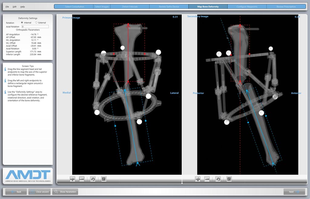
Image: Radiographic analysis softwatre helps correct complex fractures and deformities (Photo courtesy of AMDT Holdings).
An innovative software application uses radiopaque markers to automate correction of limb deformities when using a spatial frame external fixator.
The AMDT Holdings (Collierville, TN, USA) SixFix system, indicated for use with the Smith & Nephew (London, United Kingdom) Taylor Spatial Frame, consists of deformity analysis and correction software and complementary instrumentation that are designed to reduce the time and effort involved in the manual input and analysis of deformity measurement values. These values, mostly derived from patient radiographs, are required in order to build a prescription plan consisting of daily adjustments to the spatial frame fixator over a period of weeks or months.
With the SixFix system, the surgeon attaches six proprietary radiopaque fiducial markers to the rings of a spatial frame external fixator, and inputs spatial frame characteristics--ring diameter, strut length, and axial rotation--into the SixFix software. The six radiopaque markers provide full descriptive positional data to the software that enable it to create a precise three-dimensional (3-D) replica of the frame and the deformity, regardless of how the radiographic images were taken, using sophisticated algorithms that correct radiographic imperfections, such as variable magnification and perspective distortion.
The data is then used to graphically map the deformity with line segments that define the axes of the bone fragments. The SixFix software also offers options to specify waypoints, customized treatment milestones that the surgeon uses to set the order and duration of each correction. AMDT Holdings is also in the development process of a proprietary hexapod fixation device that will enhance SixFix capabilities, according to Patrick Mullaney, President and CEO of AMDT Holdings.
“Of all the advantages offered by the SixFix software, time-savings is one benefit that I will realize right away. The current devices require a great deal of time and manual effort during my clinic hours to measure and record the data necessary to describe the frame and the deformity as represented by their radiographs,” said Stephen Frania, DPM, of Foot & Ankle Specialists of Ohio (Mentor, OH, USA). “I bring these patients in weekly to evaluate the progress of their correction. The time I save and the improved accuracy when using the SixFix software is significant over these repeat visits.”
Spatial frame fixators usually consist of two rings connected by six adjustable connectors called struts. This type of construct can also be referred to as a hexapod. Fixation elements connect the rings to bone fragments; small, simultaneous changes in the lengths of multiple struts alter the alignment of the bone fragments in relation to one another. A prescription consisting of daily adjustments is used to realign or correct the misalignments, resulting is a corrected deformity and a healed fracture.
Related Links:
AMDT Holdings
Smith & Nephew
Latest General/Advanced Imaging News
- Bone Density Test Uses Existing CT Images to Predict Fractures
- AI Predicts Cardiac Risk and Mortality from Routine Chest CT Scans
- Radiation Therapy Computed Tomography Solution Boosts Imaging Accuracy
- PET Scans Reveal Hidden Inflammation in Multiple Sclerosis Patients
- Artificial Intelligence Evaluates Cardiovascular Risk from CT Scans
- New AI Method Captures Uncertainty in Medical Images
- CT Coronary Angiography Reduces Need for Invasive Tests to Diagnose Coronary Artery Disease
- Novel Blood Test Could Reduce Need for PET Imaging of Patients with Alzheimer’s
- CT-Based Deep Learning Algorithm Accurately Differentiates Benign From Malignant Vertebral Fractures
- Minimally Invasive Procedure Could Help Patients Avoid Thyroid Surgery
- Self-Driving Mobile C-Arm Reduces Imaging Time during Surgery
- AR Application Turns Medical Scans Into Holograms for Assistance in Surgical Planning
- Imaging Technology Provides Ground-Breaking New Approach for Diagnosing and Treating Bowel Cancer
- CT Coronary Calcium Scoring Predicts Heart Attacks and Strokes
- AI Model Detects 90% of Lymphatic Cancer Cases from PET and CT Images
- Breakthrough Technology Revolutionizes Breast Imaging
Channels
Radiography
view channel
Novel Breast Imaging System Proves As Effective As Mammography
Breast cancer remains the most frequently diagnosed cancer among women. It is projected that one in eight women will be diagnosed with breast cancer during her lifetime, and one in 42 women who turn 50... Read more
AI Assistance Improves Breast-Cancer Screening by Reducing False Positives
Radiologists typically detect one case of cancer for every 200 mammograms reviewed. However, these evaluations often result in false positives, leading to unnecessary patient recalls for additional testing,... Read moreMRI
view channel
Low-Cost Whole-Body MRI Device Combined with AI Generates High-Quality Results
Magnetic Resonance Imaging (MRI) has significantly transformed healthcare, providing a noninvasive, radiation-free method for detailed imaging. It is especially promising for the future of medical diagnosis... Read more
World's First Whole-Body Ultra-High Field MRI Officially Comes To Market
The world's first whole-body ultra-high field (UHF) MRI has officially come to market, marking a remarkable advancement in diagnostic radiology. United Imaging (Shanghai, China) has secured clearance from the U.... Read moreUltrasound
view channel.jpg)
Diagnostic System Automatically Analyzes TTE Images to Identify Congenital Heart Disease
Congenital heart disease (CHD) is one of the most prevalent congenital anomalies worldwide, presenting substantial health and financial challenges for affected patients. Early detection and treatment of... Read more
Super-Resolution Imaging Technique Could Improve Evaluation of Cardiac Conditions
The heart depends on efficient blood circulation to pump blood throughout the body, delivering oxygen to tissues and removing carbon dioxide and waste. Yet, when heart vessels are damaged, it can disrupt... Read more
First AI-Powered POC Ultrasound Diagnostic Solution Helps Prioritize Cases Based On Severity
Ultrasound scans are essential for identifying and diagnosing various medical conditions, but often, patients must wait weeks or months for results due to a shortage of qualified medical professionals... Read moreNuclear Medicine
view channel
New PET Biomarker Predicts Success of Immune Checkpoint Blockade Therapy
Immunotherapies, such as immune checkpoint blockade (ICB), have shown promising clinical results in treating melanoma, non-small cell lung cancer, and other tumor types. However, the effectiveness of these... Read moreNew PET Agent Rapidly and Accurately Visualizes Lesions in Clear Cell Renal Cell Carcinoma Patients
Clear cell renal cell cancer (ccRCC) represents 70-80% of renal cell carcinoma cases. While localized disease can be effectively treated with surgery and ablative therapies, one-third of patients either... Read more
New Imaging Technique Monitors Inflammation Disorders without Radiation Exposure
Imaging inflammation using traditional radiological techniques presents significant challenges, including radiation exposure, poor image quality, high costs, and invasive procedures. Now, new contrast... Read more
New SPECT/CT Technique Could Change Imaging Practices and Increase Patient Access
The development of lead-212 (212Pb)-PSMA–based targeted alpha therapy (TAT) is garnering significant interest in treating patients with metastatic castration-resistant prostate cancer. The imaging of 212Pb,... Read moreImaging IT
view channel
New Google Cloud Medical Imaging Suite Makes Imaging Healthcare Data More Accessible
Medical imaging is a critical tool used to diagnose patients, and there are billions of medical images scanned globally each year. Imaging data accounts for about 90% of all healthcare data1 and, until... Read more
Global AI in Medical Diagnostics Market to Be Driven by Demand for Image Recognition in Radiology
The global artificial intelligence (AI) in medical diagnostics market is expanding with early disease detection being one of its key applications and image recognition becoming a compelling consumer proposition... Read moreIndustry News
view channel
Hologic Acquires UK-Based Breast Surgical Guidance Company Endomagnetics Ltd.
Hologic, Inc. (Marlborough, MA, USA) has entered into a definitive agreement to acquire Endomagnetics Ltd. (Cambridge, UK), a privately held developer of breast cancer surgery technologies, for approximately... Read more
Bayer and Google Partner on New AI Product for Radiologists
Medical imaging data comprises around 90% of all healthcare data, and it is a highly complex and rich clinical data modality and serves as a vital tool for diagnosing patients. Each year, billions of medical... Read more


