


Portable Orthopedic CT Systems Image Extremities
|
By MedImaging International staff writers Posted on 16 Apr 2018 |
|
.")
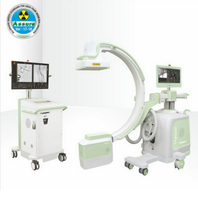
Image: The LineUP orthopedic extremity CT system (Photo courtesy of CurveBeam).
Two compact computerized tomography (CT) systems provide radiology and orthopedic specialists with three-dimensional (3D) bone detail of extremities in locations convenient to the patient.
The CurveBeam (Warrington, PA, USA) LineUP and InReach orthopedic extremity CT systems can be plugged into a standard wall outlet and have minimal shielding requirements, as the radiation dose is significantly less than that of a conventional CT scan. The LineUP system can perform bilateral scans of entire legs, from below the heel to above the knee, with an adaptive chair permitting scanning of the hand, wrist, and elbow as well. LineUP also permits the patient to stand during the scan, so that anatomy can be assessed while in a load bearing position.
The InReach system is intended for CT imaging of the hand, wrist, elbow, and lower extremities in non-weight bearing position, with the effective dose less than 2 micro Sieverts for the average adult patient for a hand scan, and 3 - 6 micro Sieverts for a standard foot/ankle scan; a Lite Dose protocol reduces radiation dose further by 56-65%. Both systems offer quick scan times of 30 seconds or less, providing 3D renderings, multi-planar slices, and X-Ray views. The images are digital imaging and communications in medicine (DICOM) and picture archiving and communication system (PACS) compliant.
“The InReach will revolutionize the speed and accuracy of assessment of upper extremity conditions that specialists have traditionally found challenging to diagnose with plain X-Ray, such as scaphoid fractures,” said Arun Singh, President and CEO of CurveBeam. “The InReach continues the company's mission to elevate advanced diagnostic imaging capabilities to enhance orthopedic care.”
“We purchased a CurveBeam InReach unit for our practice, and it has heightened the level of care that we can provide for our patients. Integration was seamless,” said Josef Zoldos, MD, DDS, of the Arizona Center for Hand Surgery (Phoenix, USA). “The convenience factor notwithstanding, the images that are generated are exemplary. The supplied software is extremely powerful and allows multidimensional image manipulation.”
Gold Member
Solid State Kv/Dose Multi-Sensor
AGMS-DM+
Ultrasound Doppler System
Doppler BT-200
New
Ultrasound Table
Powered Ultrasound Table-Flat Top
C-Arm with FPD
Digiscan V20 / V30

Latest General/Advanced Imaging News
- Artificial Intelligence Evaluates Cardiovascular Risk from CT Scans
- New AI Method Captures Uncertainty in Medical Images
- CT Coronary Angiography Reduces Need for Invasive Tests to Diagnose Coronary Artery Disease
- Novel Blood Test Could Reduce Need for PET Imaging of Patients with Alzheimer’s
- CT-Based Deep Learning Algorithm Accurately Differentiates Benign From Malignant Vertebral Fractures
- Minimally Invasive Procedure Could Help Patients Avoid Thyroid Surgery
- Self-Driving Mobile C-Arm Reduces Imaging Time during Surgery
- AR Application Turns Medical Scans Into Holograms for Assistance in Surgical Planning
- Imaging Technology Provides Ground-Breaking New Approach for Diagnosing and Treating Bowel Cancer
- CT Coronary Calcium Scoring Predicts Heart Attacks and Strokes
- AI Model Detects 90% of Lymphatic Cancer Cases from PET and CT Images
- Breakthrough Technology Revolutionizes Breast Imaging
- State-Of-The-Art System Enhances Accuracy of Image-Guided Diagnostic and Interventional Procedures
- Catheter-Based Device with New Cardiovascular Imaging Approach Offers Unprecedented View of Dangerous Plaques
- AI Model Draws Maps to Accurately Identify Tumors and Diseases in Medical Images
- AI-Enabled CT System Provides More Accurate and Reliable Imaging Results
Channels
Radiography
view channel")
Novel Breast Imaging System Proves As Effective As Mammography
Breast cancer remains the most frequently diagnosed cancer among women. It is projected that one in eight women will be diagnosed with breast cancer during her lifetime, and one in 42 women who turn 50... Read more")
AI Assistance Improves Breast-Cancer Screening by Reducing False Positives
Radiologists typically detect one case of cancer for every 200 mammograms reviewed. However, these evaluations often result in false positives, leading to unnecessary patient recalls for additional testing,... Read more")
")
MRI
view channel.jpg "Image: The emerging role of MRI alongside PSA testing is redefining prostate cancer diagnostics (Photo courtesy of 123RF)")
Combining MRI with PSA Testing Improves Clinical Outcomes for Prostate Cancer Patients
Prostate cancer is a leading health concern globally, consistently being one of the most common types of cancer among men and a major cause of cancer-related deaths. In the United States, it is the most... Read more")
PET/MRI Improves Diagnostic Accuracy for Prostate Cancer Patients
The Prostate Imaging Reporting and Data System (PI-RADS) is a five-point scale to assess potential prostate cancer in MR images. PI-RADS category 3 which offers an unclear suggestion of clinically significant... Read more")
Next Generation MR-Guided Focused Ultrasound Ushers In Future of Incisionless Neurosurgery
Essential tremor, often called familial, idiopathic, or benign tremor, leads to uncontrollable shaking that significantly affects a person’s life. When traditional medications do not alleviate symptoms,... Read more")
Two-Part MRI Scan Detects Prostate Cancer More Quickly without Compromising Diagnostic Quality
Prostate cancer ranks as the most prevalent cancer among men. Over the last decade, the introduction of MRI scans has significantly transformed the diagnosis process, marking the most substantial advancement... Read moreUltrasound
view channel.jpg "Image: The Sonic Lumen Tomography (SLT) intravascular ultrasound system has received FDA 510(k) clearance (Photo courtesy of Provisio Medical)")
Groundbreaking Technology Enables Precise, Automatic Measurement of Peripheral Blood Vessels
The current standard of care of using angiographic information is often inadequate for accurately assessing vessel size in the estimated 20 million people in the U.S. who suffer from peripheral vascular disease.... Read more")
Deep Learning Advances Super-Resolution Ultrasound Imaging
Ultrasound localization microscopy (ULM) is an advanced imaging technique that offers high-resolution visualization of microvascular structures. It employs microbubbles, FDA-approved contrast agents, injected... Read more")
Novel Ultrasound-Launched Targeted Nanoparticle Eliminates Biofilm and Bacterial Infection
Biofilms, formed by bacteria aggregating into dense communities for protection against harsh environmental conditions, are a significant contributor to various infectious diseases. Biofilms frequently... Read more")
Nuclear Medicine
view channel")
New SPECT/CT Technique Could Change Imaging Practices and Increase Patient Access
The development of lead-212 (212Pb)-PSMA–based targeted alpha therapy (TAT) is garnering significant interest in treating patients with metastatic castration-resistant prostate cancer. The imaging of 212Pb,... Read moreNew Radiotheranostic System Detects and Treats Ovarian Cancer Noninvasively
Ovarian cancer is the most lethal gynecological cancer, with less than a 30% five-year survival rate for those diagnosed in late stages. Despite surgery and platinum-based chemotherapy being the standard... Read more")
AI System Automatically and Reliably Detects Cardiac Amyloidosis Using Scintigraphy Imaging
Cardiac amyloidosis, a condition characterized by the buildup of abnormal protein deposits (amyloids) in the heart muscle, severely affects heart function and can lead to heart failure or death without... Read moreImaging IT
view channel")
New Google Cloud Medical Imaging Suite Makes Imaging Healthcare Data More Accessible
Medical imaging is a critical tool used to diagnose patients, and there are billions of medical images scanned globally each year. Imaging data accounts for about 90% of all healthcare data1 and, until... Read more
Global AI in Medical Diagnostics Market to Be Driven by Demand for Image Recognition in Radiology
The global artificial intelligence (AI) in medical diagnostics market is expanding with early disease detection being one of its key applications and image recognition becoming a compelling consumer proposition... Read more

Industry News
view channel")
Bayer and Google Partner on New AI Product for Radiologists
Medical imaging data comprises around 90% of all healthcare data, and it is a highly complex and rich clinical data modality and serves as a vital tool for diagnosing patients. Each year, billions of medical... Read more")
")
")