Varex Imaging Showcases New X-ray Components at Trade Fair
|
By MedImaging International staff writers Posted on 29 Nov 2017 |

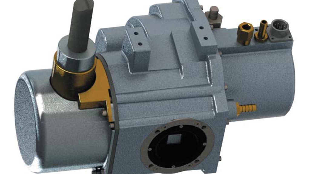
Image: The FP 1596 medical x-ray tube (Photo courtesy of Varex Imaging).
Varex Imaging Corporation (Salt Lake City, UT, USA) showcased its latest X-ray imaging tubes and sources, digital detectors, connect and control devices and software solutions at the 103rd Scientific Assembly and Annual Meeting of the Radiological Society of North America. The company also highlighted a number of other X-ray components from its product portfolio at the RSNA conference and exhibit held on November 26th – December 1st at McCormick Place in Chicago, Illinois.
Varex designs and manufactures X-ray imaging components, including tubes, digital flat panel detectors and other image processing solutions, which are key components of X-ray imaging systems. The company's X-ray sources, digital detectors, connecting devices and imaging software are used by global OEM manufacturers of X-ray imaging systems as components in their systems.
Among Varex's new X-ray imaging products on display at RSNA 2017 were its new high power, compact cardiovascular X-ray tube which incorporates a number of features to provide OEM customers with performance advantages for their cardiovascular imaging systems. Some of these advantages include high patient throughput, better imaging with lower dose, lightweight and small package size. The company also showcased its integrated CT tube sub-assembly, which combines multiple components in an optimized package that incorporates a CT tube, generator, high-voltage connector, heat exchanger and tube control unit.
At this year’s RSNA, Varex highlighted its new CMOS and amorphous-silicon based flat panel digital detectors for radiography and R&F, mammography and fluoroscopy. Additionally, the company introduced a full suite of manual and automated collimators for fixed and mobile systems, along with new dedicated software applications for planning, acquisition, and analysis. Varex also showcased its Nexus family of image acquisition workstations and image correction and 2D/3D image reconstruction software for CBCT applications in dental and medical modalities.
Latest RSNA 2017 News
- Siemens Healthineers Launches New Mammography System at RSNA
- New Real-Time Imaging AI Platform Unveiled
- Bracco Diagnostics Highlights Advancement in Diagnostic Imaging Portfolio
- Samsung Unveils New Mobile CT at RSNA 2017
- Thales Unveils World’s First Portable Detector with Embedded Patient ID
- PACSHealth Showcases DoseMonitor Upgrade at Chicago Trade Show
- Carestream Exhibits New Diagnostic Imaging Solutions in Chicago
- Siemens Healthineers Spotlights Advances in Breast Imaging
- Agfa HealthCare Presents Advances in AI and Machine Learning at RSNA 2017
- Philips Showcases New Imaging Systems and Informatics Portfolio at RSNA
- Fujifilm Debuts New FDR Go PLUS Version Portable System
- Canon Showcases New DR Systems at Medical Imaging Fair
- Aspect Imaging Presents Neonatal MRI System at RSNA 2017
- Visage Imaging Debuts AI Offerings at Chicago Show
- Carestream Joins Zebra Medical Vision to Provide Access to AI Algorithms
Channels
Radiography
view channel
Novel Breast Imaging System Proves As Effective As Mammography
Breast cancer remains the most frequently diagnosed cancer among women. It is projected that one in eight women will be diagnosed with breast cancer during her lifetime, and one in 42 women who turn 50... Read more
AI Assistance Improves Breast-Cancer Screening by Reducing False Positives
Radiologists typically detect one case of cancer for every 200 mammograms reviewed. However, these evaluations often result in false positives, leading to unnecessary patient recalls for additional testing,... Read moreMRI
view channel
Diamond Dust Could Offer New Contrast Agent Option for Future MRI Scans
Gadolinium, a heavy metal used for over three decades as a contrast agent in medical imaging, enhances the clarity of MRI scans by highlighting affected areas. Despite its utility, gadolinium not only... Read more.jpg)
Combining MRI with PSA Testing Improves Clinical Outcomes for Prostate Cancer Patients
Prostate cancer is a leading health concern globally, consistently being one of the most common types of cancer among men and a major cause of cancer-related deaths. In the United States, it is the most... Read more
PET/MRI Improves Diagnostic Accuracy for Prostate Cancer Patients
The Prostate Imaging Reporting and Data System (PI-RADS) is a five-point scale to assess potential prostate cancer in MR images. PI-RADS category 3 which offers an unclear suggestion of clinically significant... Read more
Next Generation MR-Guided Focused Ultrasound Ushers In Future of Incisionless Neurosurgery
Essential tremor, often called familial, idiopathic, or benign tremor, leads to uncontrollable shaking that significantly affects a person’s life. When traditional medications do not alleviate symptoms,... Read moreUltrasound
view channel.jpg)
Groundbreaking Technology Enables Precise, Automatic Measurement of Peripheral Blood Vessels
The current standard of care of using angiographic information is often inadequate for accurately assessing vessel size in the estimated 20 million people in the U.S. who suffer from peripheral vascular disease.... Read more
Deep Learning Advances Super-Resolution Ultrasound Imaging
Ultrasound localization microscopy (ULM) is an advanced imaging technique that offers high-resolution visualization of microvascular structures. It employs microbubbles, FDA-approved contrast agents, injected... Read more
Novel Ultrasound-Launched Targeted Nanoparticle Eliminates Biofilm and Bacterial Infection
Biofilms, formed by bacteria aggregating into dense communities for protection against harsh environmental conditions, are a significant contributor to various infectious diseases. Biofilms frequently... Read moreNuclear Medicine
view channel
New Imaging Technique Monitors Inflammation Disorders without Radiation Exposure
Imaging inflammation using traditional radiological techniques presents significant challenges, including radiation exposure, poor image quality, high costs, and invasive procedures. Now, new contrast... Read more
New SPECT/CT Technique Could Change Imaging Practices and Increase Patient Access
The development of lead-212 (212Pb)-PSMA–based targeted alpha therapy (TAT) is garnering significant interest in treating patients with metastatic castration-resistant prostate cancer. The imaging of 212Pb,... Read moreNew Radiotheranostic System Detects and Treats Ovarian Cancer Noninvasively
Ovarian cancer is the most lethal gynecological cancer, with less than a 30% five-year survival rate for those diagnosed in late stages. Despite surgery and platinum-based chemotherapy being the standard... Read more
AI System Automatically and Reliably Detects Cardiac Amyloidosis Using Scintigraphy Imaging
Cardiac amyloidosis, a condition characterized by the buildup of abnormal protein deposits (amyloids) in the heart muscle, severely affects heart function and can lead to heart failure or death without... Read moreGeneral/Advanced Imaging
view channel
PET Scans Reveal Hidden Inflammation in Multiple Sclerosis Patients
A key challenge for clinicians treating patients with multiple sclerosis (MS) is that after a certain amount of time, they continue to worsen even though their MRIs show no change. A new study has now... Read more
Artificial Intelligence Evaluates Cardiovascular Risk from CT Scans
Chest computed tomography (CT) is a common diagnostic tool, with approximately 15 million scans conducted each year in the United States, though many are underutilized or not fully explored.... Read more
New AI Method Captures Uncertainty in Medical Images
In the field of biomedicine, segmentation is the process of annotating pixels from an important structure in medical images, such as organs or cells. Artificial Intelligence (AI) models are utilized to... Read more.jpg)
CT Coronary Angiography Reduces Need for Invasive Tests to Diagnose Coronary Artery Disease
Coronary artery disease (CAD), one of the leading causes of death worldwide, involves the narrowing of coronary arteries due to atherosclerosis, resulting in insufficient blood flow to the heart muscle.... Read moreImaging IT
view channel
New Google Cloud Medical Imaging Suite Makes Imaging Healthcare Data More Accessible
Medical imaging is a critical tool used to diagnose patients, and there are billions of medical images scanned globally each year. Imaging data accounts for about 90% of all healthcare data1 and, until... Read more
Global AI in Medical Diagnostics Market to Be Driven by Demand for Image Recognition in Radiology
The global artificial intelligence (AI) in medical diagnostics market is expanding with early disease detection being one of its key applications and image recognition becoming a compelling consumer proposition... Read moreIndustry News
view channel
Bayer and Google Partner on New AI Product for Radiologists
Medical imaging data comprises around 90% of all healthcare data, and it is a highly complex and rich clinical data modality and serves as a vital tool for diagnosing patients. Each year, billions of medical... Read more


