Photoacoustic Tomography Helps Detect Deep Cancer Cells
|
By MedImaging International staff writers Posted on 16 Nov 2015 |

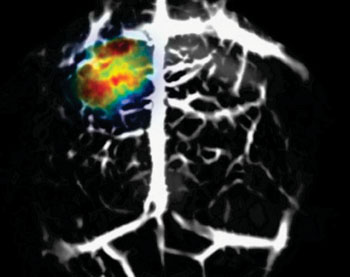
Image: Deep-tissue in vivo PAT genetic imaging of reversibly switchable BphP1 in a mouse brain (Photo courtesy of WUSTL).
Genetically modifying glioblastoma cells with a bacterial phytochrome enables them to be identified as deep as one centimeter inside tissue using photoacoustic tomography (PAT).
Researchers at Washington University (WUSTL; St. Louis, MO, USA) and Albert Einstein College of Medicine (New York, NY, USA) successfully combined deep-penetration, high-resolution PAT with a BphP1, a reversibly switchable, phytochrome derived from Rhodopseudomonas palustris bacterium. Since BphP1 has the ability to differentiate between red and near-infrared (NIR) light-absorption states, the researchers took images of the modified cancerous tissue using the two types of light and compared them to get a highly sensitive, high-resolution image of the cancer cells.
Furthermore, the combined single-wavelength PAT and efficient BphP1 photo-switching between red and NIR states enabled differential imaging with substantially decreased background signals, enhanced detection sensitivity, increased penetration depth, and improved spatial resolution. This allowed the researchers to monitor tumor growth and metastasis with 100-μm resolution at depths approaching 10 mm and image individual cancer cells with a sub-optical diffraction resolution of about 140 nm. The study was published on November 9, 2015, in Nature Methods.
“This technique is extremely useful for cancer imaging. When we first look at the early-stage cancer, cancer tissue does not differ much from the background, healthy tissue; because the abundant blood gives strong signals, the cancer cells don't stand out,” said lead author biomedical engineer Junjie Yao, PhD, of WUSTL. “Now, with this new technology, we can see cancer cells in a tiny bit of tissue when the cancer is small.”
“Genetic encoding of the protein allows us to image and track targeted biological processes deep in tissue. The optical switching property of the protein enables new imaging capability,” said senior author Lihong Wang, PhD, of WUSTL. “This technology provides a promising new tool to biologists for high-resolution, deep imaging of cancer with genetic specificity as well as for drug screening in living tissue.”
PAT of genetically encoded probes allows for imaging of targeted biological processes deep in tissues with high spatial resolution; however, high background signals from blood can limit the achievable detection sensitivity.
Related Links:
Washington University
Albert Einstein College of Medicine
Latest General/Advanced Imaging News
- Radiation Therapy Computed Tomography Solution Boosts Imaging Accuracy
- PET Scans Reveal Hidden Inflammation in Multiple Sclerosis Patients
- Artificial Intelligence Evaluates Cardiovascular Risk from CT Scans
- New AI Method Captures Uncertainty in Medical Images
- CT Coronary Angiography Reduces Need for Invasive Tests to Diagnose Coronary Artery Disease
- Novel Blood Test Could Reduce Need for PET Imaging of Patients with Alzheimer’s
- CT-Based Deep Learning Algorithm Accurately Differentiates Benign From Malignant Vertebral Fractures
- Minimally Invasive Procedure Could Help Patients Avoid Thyroid Surgery
- Self-Driving Mobile C-Arm Reduces Imaging Time during Surgery
- AR Application Turns Medical Scans Into Holograms for Assistance in Surgical Planning
- Imaging Technology Provides Ground-Breaking New Approach for Diagnosing and Treating Bowel Cancer
- CT Coronary Calcium Scoring Predicts Heart Attacks and Strokes
- AI Model Detects 90% of Lymphatic Cancer Cases from PET and CT Images
- Breakthrough Technology Revolutionizes Breast Imaging
- State-Of-The-Art System Enhances Accuracy of Image-Guided Diagnostic and Interventional Procedures
- Catheter-Based Device with New Cardiovascular Imaging Approach Offers Unprecedented View of Dangerous Plaques
Channels
Radiography
view channel
Novel Breast Imaging System Proves As Effective As Mammography
Breast cancer remains the most frequently diagnosed cancer among women. It is projected that one in eight women will be diagnosed with breast cancer during her lifetime, and one in 42 women who turn 50... Read more
AI Assistance Improves Breast-Cancer Screening by Reducing False Positives
Radiologists typically detect one case of cancer for every 200 mammograms reviewed. However, these evaluations often result in false positives, leading to unnecessary patient recalls for additional testing,... Read moreMRI
view channel
Low-Cost Whole-Body MRI Device Combined with AI Generates High-Quality Results
Magnetic Resonance Imaging (MRI) has significantly transformed healthcare, providing a noninvasive, radiation-free method for detailed imaging. It is especially promising for the future of medical diagnosis... Read more
World's First Whole-Body Ultra-High Field MRI Officially Comes To Market
The world's first whole-body ultra-high field (UHF) MRI has officially come to market, marking a remarkable advancement in diagnostic radiology. United Imaging (Shanghai, China) has secured clearance from the U.... Read moreUltrasound
view channel
First AI-Powered POC Ultrasound Diagnostic Solution Helps Prioritize Cases Based On Severity
Ultrasound scans are essential for identifying and diagnosing various medical conditions, but often, patients must wait weeks or months for results due to a shortage of qualified medical professionals... Read more
Largest Model Trained On Echocardiography Images Assesses Heart Structure and Function
Foundation models represent an exciting frontier in generative artificial intelligence (AI), yet many lack the specialized medical data needed to make them applicable in healthcare settings.... Read more.jpg)
Groundbreaking Technology Enables Precise, Automatic Measurement of Peripheral Blood Vessels
The current standard of care of using angiographic information is often inadequate for accurately assessing vessel size in the estimated 20 million people in the U.S. who suffer from peripheral vascular disease.... Read moreNuclear Medicine
view channelNew PET Agent Rapidly and Accurately Visualizes Lesions in Clear Cell Renal Cell Carcinoma Patients
Clear cell renal cell cancer (ccRCC) represents 70-80% of renal cell carcinoma cases. While localized disease can be effectively treated with surgery and ablative therapies, one-third of patients either... Read more
New Imaging Technique Monitors Inflammation Disorders without Radiation Exposure
Imaging inflammation using traditional radiological techniques presents significant challenges, including radiation exposure, poor image quality, high costs, and invasive procedures. Now, new contrast... Read more
New SPECT/CT Technique Could Change Imaging Practices and Increase Patient Access
The development of lead-212 (212Pb)-PSMA–based targeted alpha therapy (TAT) is garnering significant interest in treating patients with metastatic castration-resistant prostate cancer. The imaging of 212Pb,... Read moreImaging IT
view channel
New Google Cloud Medical Imaging Suite Makes Imaging Healthcare Data More Accessible
Medical imaging is a critical tool used to diagnose patients, and there are billions of medical images scanned globally each year. Imaging data accounts for about 90% of all healthcare data1 and, until... Read more
Global AI in Medical Diagnostics Market to Be Driven by Demand for Image Recognition in Radiology
The global artificial intelligence (AI) in medical diagnostics market is expanding with early disease detection being one of its key applications and image recognition becoming a compelling consumer proposition... Read moreIndustry News
view channel
Hologic Acquires UK-Based Breast Surgical Guidance Company Endomagnetics Ltd.
Hologic, Inc. (Marlborough, MA, USA) has entered into a definitive agreement to acquire Endomagnetics Ltd. (Cambridge, UK), a privately held developer of breast cancer surgery technologies, for approximately... Read more
Bayer and Google Partner on New AI Product for Radiologists
Medical imaging data comprises around 90% of all healthcare data, and it is a highly complex and rich clinical data modality and serves as a vital tool for diagnosing patients. Each year, billions of medical... Read more


