MRI-Based Machine Learning System Diagnoses Alzheimer’s with Single Brain Scan
|
By MedImaging International staff writers Posted on 21 Jun 2022 |

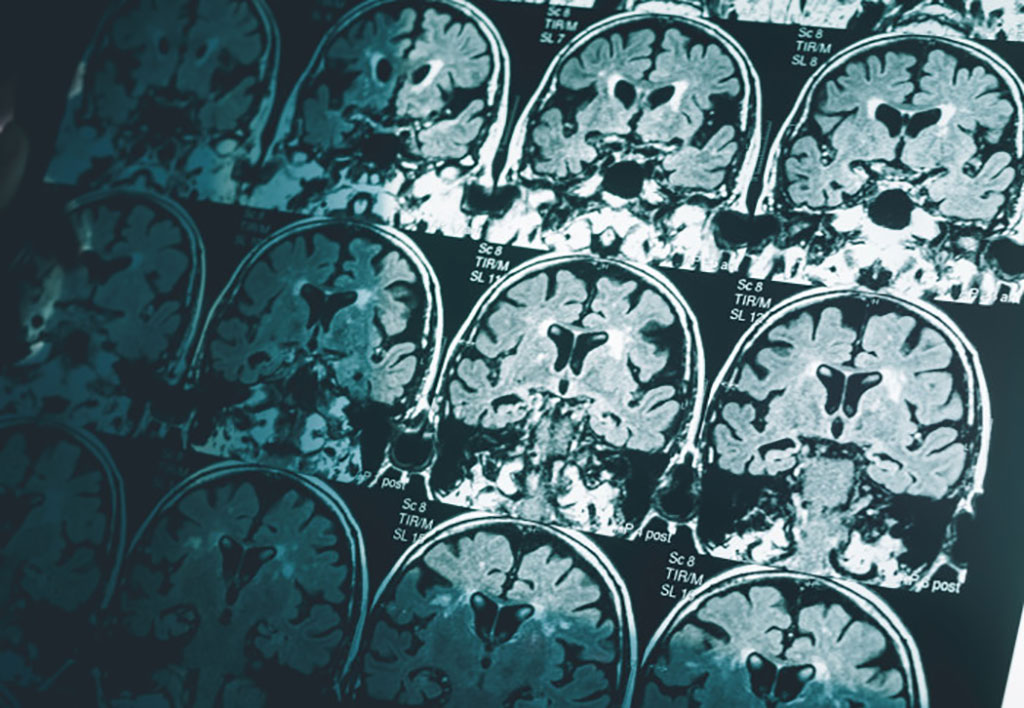
Alzheimer’s disease is the most common form of dementia, and although most people with Alzheimer’s develop it after the age of 65, people under this age can develop it too. The most frequent symptoms of dementia are memory loss and difficulties with thinking, problem solving and language. Doctors currently use a raft of tests to diagnose Alzheimer’s disease, including memory and cognitive tests and brain scans. The scans are used to check for protein deposits in the brain and shrinkage of the hippocampus, the area of the brain linked to memory. All of these tests can take several weeks, both to arrange and to process. Now, a new approach requires just one of these – a magnetic resonance imaging (MRI) brain scan taken on a standard 1.5 Tesla machine, which is commonly found in most hospitals.
Researchers at the Imperial College London (London, UK) used machine learning technology to look at structural features within the brain, including in regions not previously associated with Alzheimer’s. The advantage of the technique is its simplicity and the fact that it can identify the disease at an early stage when it can be very difficult to diagnose. Although there is no cure for Alzheimer’s disease, getting a diagnosis quickly at an early stage helps patients. It allows them to access help and support, get treatment to manage their symptoms and plan for the future. Being able to accurately identify patients at an early stage of the disease will also help researchers to understand the brain changes that trigger the disease, and support development and trials of new treatments.
The researchers adapted an algorithm developed for use in classifying cancer tumors, and applied it to the brain. They divided the brain into 115 regions and allocated 660 different features, such as size, shape and texture, to assess each region. They then trained the algorithm to identify where changes to these features could accurately predict the existence of Alzheimer’s disease. Using data from the Alzheimer’s Disease Neuroimaging Initiative, the team tested their approach on brain scans from over 400 patients with early and later stage Alzheimer’s, healthy controls and patients with other neurological conditions, including frontotemporal dementia and Parkinson’s disease. They also tested it with data from over 80 patients undergoing diagnostic tests for Alzheimer’s at Imperial College Healthcare NHS Trust.
The researchers found that in 98% of cases, the MRI-based machine learning system alone could accurately predict whether the patient had Alzheimer’s disease or not. It was also able to distinguish between early and late-stage Alzheimer’s with fairly high accuracy, in 79 per cent of patients. The new system also spotted changes in areas of the brain not previously associated with Alzheimer’s disease, including the cerebellum (the part of the brain that coordinates and regulates physical activity) and the ventral diencephalon (linked to the senses, sight and hearing). This opens up potential new avenues for research into these areas and their links to Alzheimer’s disease.
“Currently no other simple and widely available methods can predict Alzheimer’s disease with this level of accuracy, so our research is an important step forward,” said Professor Eric Aboagye, from Imperial’s Department of Surgery and Cancer, who led the research. “Many patients who present with Alzheimer’s at memory clinics do also have other neurological conditions, but even within this group our system could pick out those patients who had Alzheimer’s from those who did not.”
“Waiting for a diagnosis can be a horrible experience for patients and their families,” added Professor Aboagye. “If we could cut down the amount of time they have to wait, make diagnosis a simpler process, and reduce some of the uncertainty, that would help a great deal. Our new approach could also identify early-stage patients for clinical trials of new drug treatments or lifestyle changes, which is currently very hard to do.”
“Although neuroradiologists already interpret MRI scans to help diagnose Alzheimer’s, there are likely to be features of the scans that aren’t visible, even to specialists,” explained Dr. Paresh Malhotra, a consultant neurologist at Imperial College Healthcare NHS Trust and researcher in Imperial’s Department of Brain Sciences. “Using an algorithm able to select texture and subtle structural features in the brain that are affected by Alzheimer’s could really enhance the information we can gain from standard imaging techniques.”
Related Links:
Imperial College London
Latest MRI News
- AI Model Reads and Diagnoses Brain MRI in Seconds
- MRI Scan Breakthrough to Help Avoid Risky Invasive Tests for Heart Patients
- MRI Scans Reveal Signature Patterns of Brain Activity to Predict Recovery from TBI
- Novel Imaging Approach to Improve Treatment for Spinal Cord Injuries
- AI-Assisted Model Enhances MRI Heart Scans
- AI Model Outperforms Doctors at Identifying Patients Most At-Risk of Cardiac Arrest
- New MRI Technique Reveals Hidden Heart Issues
- Shorter MRI Exam Effectively Detects Cancer in Dense Breasts
- MRI to Replace Painful Spinal Tap for Faster MS Diagnosis
- MRI Scans Can Identify Cardiovascular Disease Ten Years in Advance
- Simple Brain Scan Diagnoses Parkinson's Disease Years Before It Becomes Untreatable
- Cutting-Edge MRI Technology to Revolutionize Diagnosis of Common Heart Problem
- New MRI Technique Reveals True Heart Age to Prevent Attacks and Strokes
- AI Tool Predicts Relapse of Pediatric Brain Cancer from Brain MRI Scans
- AI Tool Tracks Effectiveness of Multiple Sclerosis Treatments Using Brain MRI Scans
- Ultra-Powerful MRI Scans Enable Life-Changing Surgery in Treatment-Resistant Epileptic Patients
Channels
Radiography
view channel
Routine Mammograms Could Predict Future Cardiovascular Disease in Women
Mammograms are widely used to screen for breast cancer, but they may also contain overlooked clues about cardiovascular health. Calcium deposits in the arteries of the breast signal stiffening blood vessels,... Read more
AI Detects Early Signs of Aging from Chest X-Rays
Chronological age does not always reflect how fast the body is truly aging, and current biological age tests often rely on DNA-based markers that may miss early organ-level decline. Detecting subtle, age-related... Read moreUltrasound
view channel
AI Model Accurately Detects Placenta Accreta in Pregnancy Before Delivery
Placenta accreta spectrum (PAS) is a life-threatening pregnancy complication in which the placenta abnormally attaches to the uterine wall. The condition is a leading cause of maternal mortality and morbidity... Read more
Portable Ultrasound Sensor to Enable Earlier Breast Cancer Detection
Breast cancer screening relies heavily on annual mammograms, but aggressive tumors can develop between scans, accounting for up to 30 percent of cases. These interval cancers are often diagnosed later,... Read more
Portable Imaging Scanner to Diagnose Lymphatic Disease in Real Time
Lymphatic disorders affect hundreds of millions of people worldwide and are linked to conditions ranging from limb swelling and organ dysfunction to birth defects and cancer-related complications.... Read more
Imaging Technique Generates Simultaneous 3D Color Images of Soft-Tissue Structure and Vasculature
Medical imaging tools often force clinicians to choose between speed, structural detail, and functional insight. Ultrasound is fast and affordable but typically limited to two-dimensional anatomy, while... Read moreNuclear Medicine
view channel
Radiopharmaceutical Molecule Marker to Improve Choice of Bladder Cancer Therapies
Targeted cancer therapies only work when tumor cells express the specific molecular structures they are designed to attack. In urothelial carcinoma, a common form of bladder cancer, the cell surface protein... Read more
Cancer “Flashlight” Shows Who Can Benefit from Targeted Treatments
Targeted cancer therapies can be highly effective, but only when a patient’s tumor expresses the specific protein the treatment is designed to attack. Determining this usually requires biopsies or advanced... Read moreGeneral/Advanced Imaging
view channel
AI Tool Offers Prognosis for Patients with Head and Neck Cancer
Oropharyngeal cancer is a form of head and neck cancer that can spread through lymph nodes, significantly affecting survival and treatment decisions. Current therapies often involve combinations of surgery,... Read more
New 3D Imaging System Addresses MRI, CT and Ultrasound Limitations
Medical imaging is central to diagnosing and managing injuries, cancer, infections, and chronic diseases, yet existing tools each come with trade-offs. Ultrasound, X-ray, CT, and MRI can be costly, time-consuming,... Read moreImaging IT
view channel
New Google Cloud Medical Imaging Suite Makes Imaging Healthcare Data More Accessible
Medical imaging is a critical tool used to diagnose patients, and there are billions of medical images scanned globally each year. Imaging data accounts for about 90% of all healthcare data1 and, until... Read more
Global AI in Medical Diagnostics Market to Be Driven by Demand for Image Recognition in Radiology
The global artificial intelligence (AI) in medical diagnostics market is expanding with early disease detection being one of its key applications and image recognition becoming a compelling consumer proposition... Read moreIndustry News
view channel
Nuclear Medicine Set for Continued Growth Driven by Demand for Precision Diagnostics
Clinical imaging services face rising demand for precise molecular diagnostics and targeted radiopharmaceutical therapy as cancer and chronic disease rates climb. A new market analysis projects rapid expansion... Read more


