Global Digital X-Ray Market to Surpass USD 14 Billion by 2026
|
By MedImaging International staff writers Posted on 13 Oct 2021 |


|

The global market for digital X-ray is projected to grow at a CAGR of more than 7% from around USD 9 billion in 2020 to over USD 14 billion by 2026, amidst all the opportunities and challenges in a significantly transformed post COVID-19 marketplace.
These are the latest findings of a new market study published by Global Industry Analysts Inc. (San Jose, CA, USA), a market research company.
Based on application, the Orthopedic segment is projected to grow at a CAGR of 8.3% to reach USD 3.3 billion in 2026 while the Cardiovascular segment is expected to register a CAGR of 7.5% for the next seven-year period. The Fluoroscopy segment is estimated to register a CAGR of 6.2% to reach USD 1.4 billion by the year 2026, led mainly by the regional markets of the US, Canada, Japan, China and Europe. These regional markets which accounted for a combined market size of USD 0.68 billion in the year 2020 are projected to reach USD 1 billion by the close of the analysis period.
China, the world`s second largest economy, will remain among the fastest growing in this cluster of regional markets with a CAGR of 11.1% over the analysis period to reach USD 3 billion by 2026. The US digital X-ray market is estimated to be worth USD 2.7 billion in 2021. Among the other noteworthy geographical markets for digital X-ray are Japan and Canada which are forecasted to register a CAGR of 3.9% and 6.5%, respectively over the analysis period. Within Europe, Germany is projected to grow at a CAGR of approximately 4.7% for the next seven-year period.
Related Links:
Global Industry Analysts Inc.









Latest Industry News News
- GE HealthCare Showcases AI-Enabled Nuclear Medicine Portfolio at SNMMI 2026
- GE HealthCare Highlights AI-Supported Radiation Therapy Tools at ESTRO 2026
- Nuclear Medicine Set for Continued Growth Driven by Demand for Precision Diagnostics
- GE HealthCare and NVIDIA Collaboration to Reimagine Diagnostic Imaging
- Patient-Specific 3D-Printed Phantoms Transform CT Imaging
- Siemens and Sectra Collaborate on Enhancing Radiology Workflows
- Bracco Diagnostics and ColoWatch Partner to Expand Availability CRC Screening Tests Using Virtual Colonoscopy
- Mindray Partners with TeleRay to Streamline Ultrasound Delivery
- Philips and Medtronic Partner on Stroke Care
- Siemens and Medtronic Enter into Global Partnership for Advancing Spine Care Imaging Technologies
- RSNA 2024 Technical Exhibits to Showcase Latest Advances in Radiology
- Bracco Collaborates with Arrayus on Microbubble-Assisted Focused Ultrasound Therapy for Pancreatic Cancer
- Innovative Collaboration to Enhance Ischemic Stroke Detection and Elevate Standards in Diagnostic Imaging
- RSNA 2024 Registration Opens
- Microsoft collaborates with Leading Academic Medical Systems to Advance AI in Medical Imaging
- GE HealthCare Acquires Intelligent Ultrasound Group’s Clinical Artificial Intelligence Business
Channels
Radiography
view channel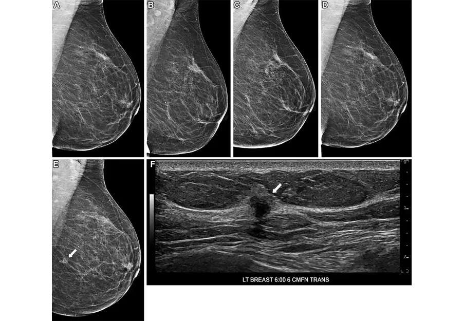
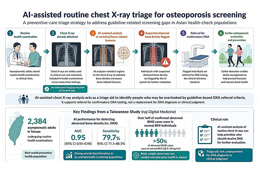
AI Tool Predicts Five-Year Breast Cancer Risk from Mammograms
Breast cancer risk assessment during routine screening is difficult because many women who develop the disease have no known genetic mutations or family history. Static risk tools provide limited discrimination... Read more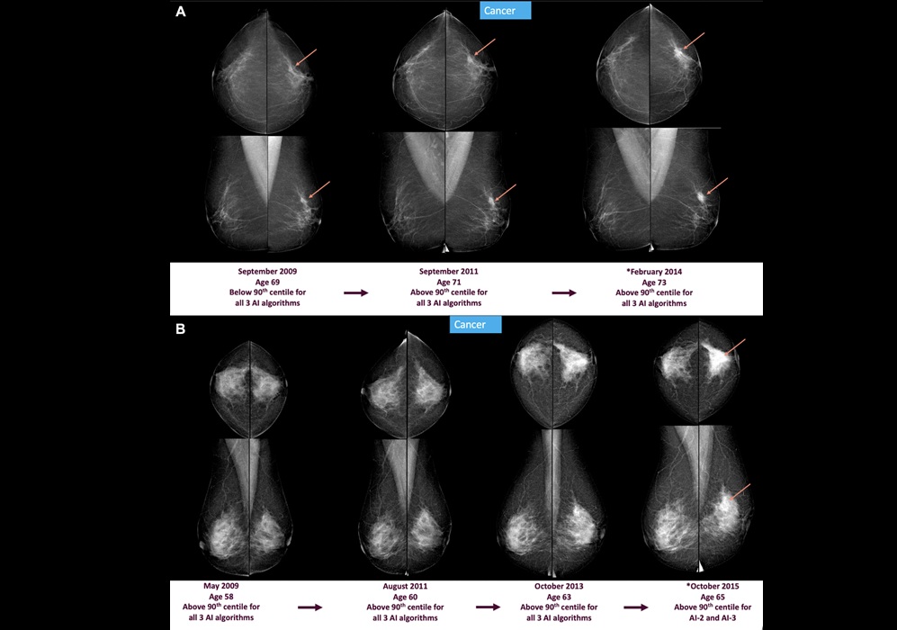
AI Mammography Tools Detect Early Breast Cancer Signs Years Before Diagnosis
Breast cancer screening aims to detect tumors before symptoms develop, but subtle mammographic changes can appear years before diagnosis and may be missed during routine reads. Delayed detection can lead... Read more
MRI
view channel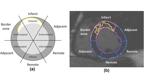
Cardiac MRI Detects Hidden Heart Dysfunction After Heart Attack
Myocardial infarction often leaves patients with persistent ventricular dysfunction that can progress without obvious signs. Detecting early decline remains difficult because standard measures may miss... Read more
AI Tool Reveals Cortical Lesions on Standard MRI in Multiple Sclerosis
Multiple sclerosis (MS) causes immune-mediated damage in the brain that leads to disability and cognitive decline. Clinicians have long recognized that lesions in the brain’s gray matter, or cortex, are... Read more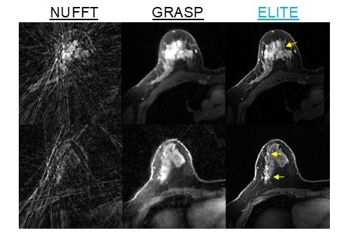
Ultrasound
view channel
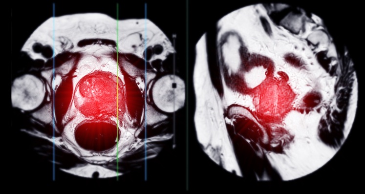
Simple 2D Ultrasound Tool Accurately Estimates Placental Volume
Stillbirth remains a major cause of perinatal loss, with about 21,000 cases annually in the United States. Although placental abnormalities are linked to adverse pregnancy outcomes, routine monitoring... Read more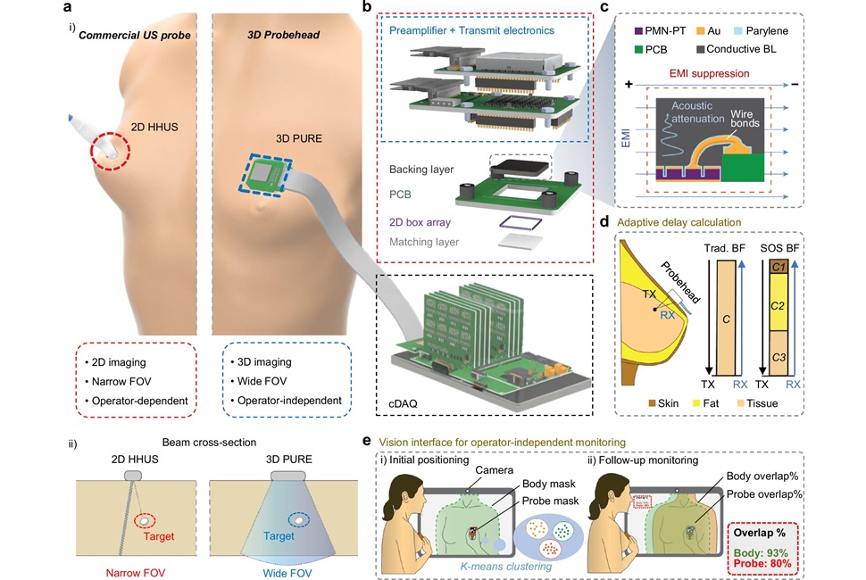
Portable 3D Ultrasound System Enables Reproducible Breast Cancer Monitoring
Breast cancer can develop between annual mammograms, and these interval cancers account for 20% to 30% of cases and tend to be more aggressive. The challenge is pronounced in people with dense breast tissue.... Read more
Nuclear Medicine
view channel
Second PSMA PET Identifies Disease in More Than Half of Negative Cases
Biochemical recurrence of prostate cancer after prostatectomy or radiation can be difficult to localize when imaging is negative. Uncertain disease sites may delay salvage therapy and expose patients to... Read more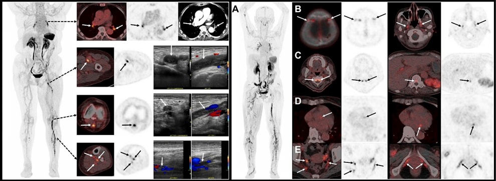
New PET Tracer Detects DVT and Pulmonary Embolism in One Scan
Deep vein thrombosis is the formation of clots in deep leg veins that can migrate to the lungs as pulmonary embolism. Rapid confirmation across both regions often requires multiple tests and can delay treatment.... Read more![Image: (A-C) Representative whole-body maximum-intensity projection images and regional fused PET/CT images from three histologically confirmed osteosarcoma patients who underwent paired [68Ga]Ga-B7-H3-BCH PET/CT and 18F-FDGE PET/CT within 7 days. (D) Multimodal imaging evaluation of patient 3, including X-ray, MRI (T2-weighted imaging, T2WI), CT, and B7-H3 PET/CT. (Photo courtesy of SNMMI)](https://globetechcdn.com/mobile_medicalimaging/images/stories/articles/article_images/2026-06-07/294811110_image_1.jpg "Image: (A-C) Representative whole-body maximum-intensity projection images and regional fused PET/CT images from three histologically confirmed osteosarcoma patients who underwent paired [68Ga]Ga-B7-H3-BCH PET/CT and 18F-FDGE PET/CT within 7 days. (D) Multimodal imaging evaluation of patient 3, including X-ray, MRI (T2-weighted imaging, T2WI), CT, and B7-H3 PET/CT. (Photo courtesy of SNMMI)")
General/Advanced Imaging
view channel
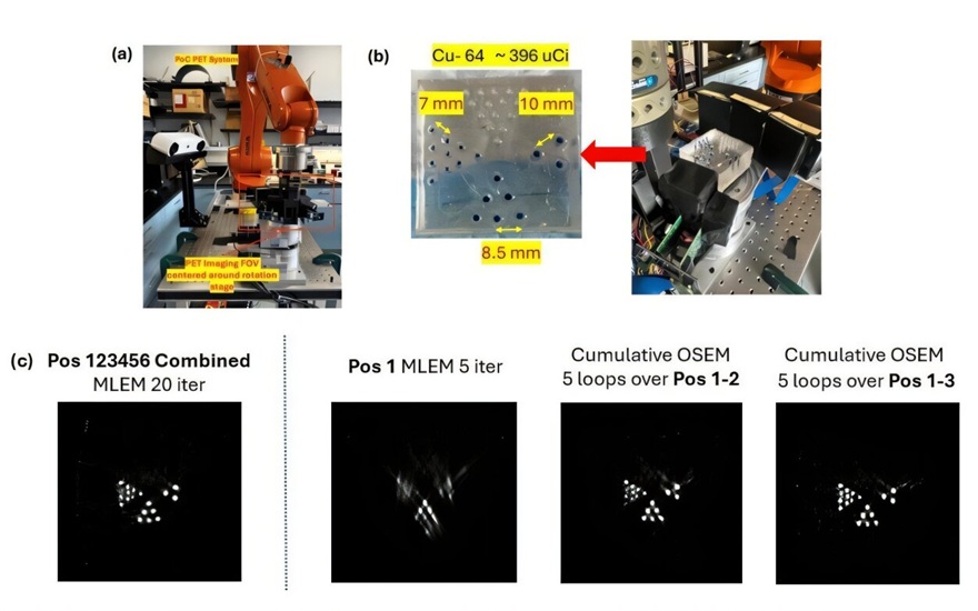
Virtual Staining Technique Creates Histology Images from CT Data
Pulmonary hypertension, a disorder marked by pathological remodeling of the pulmonary vessels, often requires detailed histologic assessment. Yet routine pathology remains anchored in labor‑intensive,... Read more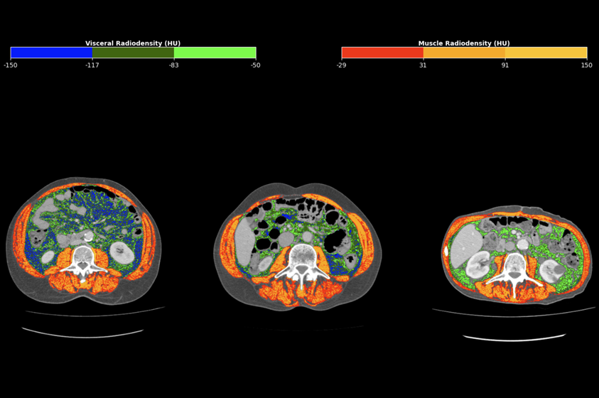
CT-Derived Biomarker Predicts Outcomes in Gastric Cancer
Gastric cancer, also known as stomach cancer, is the fifth most common malignancy worldwide and often shows heterogeneous outcomes even within the same stage. Prognostic estimates typically rely on tumor-centric... Read more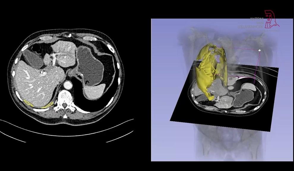
AI Tool Enhances Response Assessment and Survival Prediction in Pleural Mesothelioma
Pleural mesothelioma, a cancer that grows as a thin, irregular layer along the lung wall, is difficult to measure on imaging. Clinicians rely on diameter-based Response Evaluation Criteria in Solid Tumors... Read more
Imaging IT
view channel
Ambient AI Reporting Platform Streamlines Radiology Reporting
Radiology departments face growing imaging volumes and staffing shortages, creating reporting bottlenecks and pressure to maintain turnaround times. Conventional dictation tools document findings after... Read more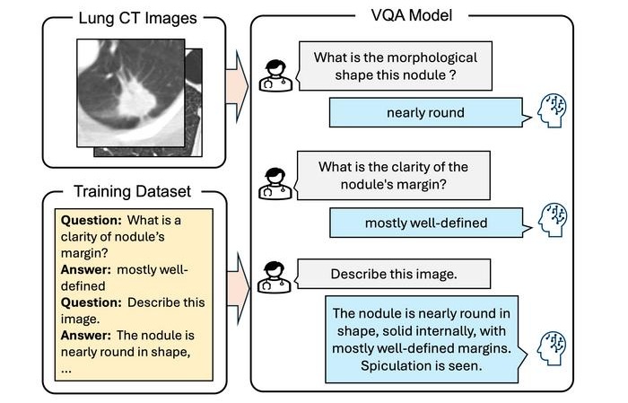
Interactive AI Tool Supports Explainable Lung Nodule Assessment
Lung cancer is a leading cause of cancer mortality, and timely characterization of pulmonary nodules on chest computed tomography (CT) is essential for directing care. Interpreting nodule morphology demands... Read more