Myelin Volume Measurement Tool Receives CE Mark
|
By MedImaging International staff writers Posted on 25 Apr 2017 |
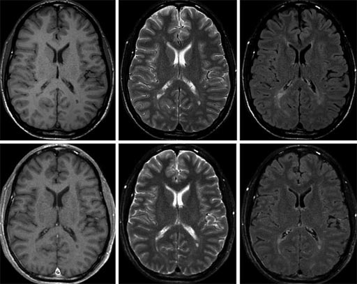
Image: The top images show conventional 1.5T MRI exams, and the lower images show SyMRI exams synthesized in a single scan (Photo courtesy of SyntheticMR).
A tool that can be used for rapid quantification of the estimated myelin volume in the brain for diagnostic imaging has been awarded the CE-mark (Conformité Européenne) for clinical use in Europe.
According to the developer of the post-processing software, the tool is compatible with Magnetic Resonance (MR) scanners from leading vendors.
The Rapid Estimation of Myelin for Diagnostic Imaging (REMyDI) tool is part of the SyntheticMR SyMRI NEURO REMyDI post-processing software package. Myelin quantification enables clinicians to monitor myelin degeneration in patients with neurodegenerative disorders, and diseases that cause brain demyelination. Clinicians can also use the tool to follow myelination in the developing brain. The new REMyDI feature will provide clinicians with automatic myelin volume measurement using data from a single 5-6-minute quantitative MRI scan. Post-processing is automatic and takes less than 10 seconds to complete.
The SyMRI software package enables clinicians to capture multiple image contrasts in one MR scan. The contrast can be adjusted following the scan to create additional images. The system provides automatic segmentation of brain tissue for objective decision support based on quantitative data. Scanner settings can be matched between scans to enable accurate comparison between exams. The images can also be matched with previous images, and conventional protocols. The SyMRI software package is CE-marked, and is pending 510(k) approval from the US Food and Drug Administration (FDA).
Latest MRI News
- AI-Assisted Model Enhances MRI Heart Scans
- AI Model Outperforms Doctors at Identifying Patients Most At-Risk of Cardiac Arrest
- New MRI Technique Reveals Hidden Heart Issues
- Shorter MRI Exam Effectively Detects Cancer in Dense Breasts
- MRI to Replace Painful Spinal Tap for Faster MS Diagnosis
- MRI Scans Can Identify Cardiovascular Disease Ten Years in Advance
- Simple Brain Scan Diagnoses Parkinson's Disease Years Before It Becomes Untreatable
- Cutting-Edge MRI Technology to Revolutionize Diagnosis of Common Heart Problem
- New MRI Technique Reveals True Heart Age to Prevent Attacks and Strokes
- AI Tool Predicts Relapse of Pediatric Brain Cancer from Brain MRI Scans
- AI Tool Tracks Effectiveness of Multiple Sclerosis Treatments Using Brain MRI Scans
- Ultra-Powerful MRI Scans Enable Life-Changing Surgery in Treatment-Resistant Epileptic Patients
- AI-Powered MRI Technology Improves Parkinson’s Diagnoses
- Biparametric MRI Combined with AI Enhances Detection of Clinically Significant Prostate Cancer
- First-Of-Its-Kind AI-Driven Brain Imaging Platform to Better Guide Stroke Treatment Options
- New Model Improves Comparison of MRIs Taken at Different Institutions
Channels
Radiography
view channel
RSNA AI Challenge Models Can Independently Interpret Mammograms
Breast cancer screening aims to detect cancers early while minimizing unnecessary recalls, yet balancing sensitivity and specificity remains a challenge. Automating detection in mammograms could help radiologists... Read more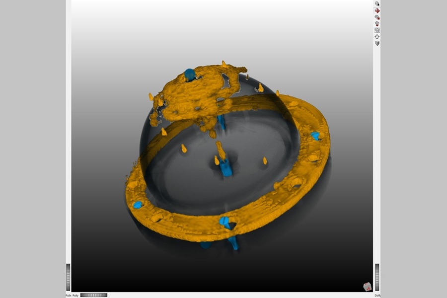
New Technique Combines X-Ray Imaging and Radar for Safer Cancer Diagnosis
X-ray imaging methods, such as mammography and computed tomography (CT) scans, are essential for diagnosing and monitoring breast and lung cancer. While CT provides valuable three-dimensional insights,... Read moreUltrasound
view channel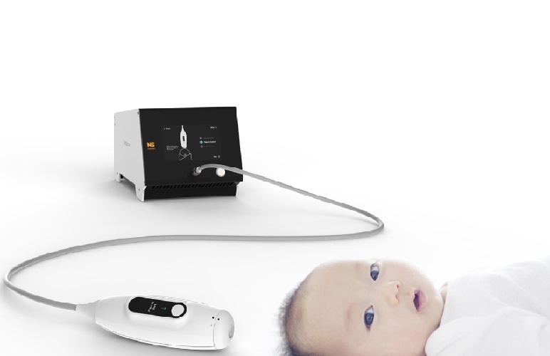
Non-Invasive Ultrasound-Based Tool Accurately Detects Infant Meningitis
Meningitis, an inflammation of the membranes surrounding the brain and spinal cord, can be fatal in infants if not diagnosed and treated early. Even when treated, it may leave lasting damage, such as cognitive... Read more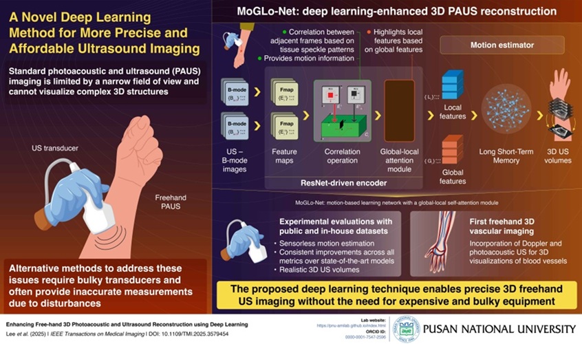
Breakthrough Deep Learning Model Enhances Handheld 3D Medical Imaging
Ultrasound imaging is a vital diagnostic technique used to visualize internal organs and tissues in real time and to guide procedures such as biopsies and injections. When paired with photoacoustic imaging... Read moreNuclear Medicine
view channel
Novel Bacteria-Specific PET Imaging Approach Detects Hard-To-Diagnose Lung Infections
Mycobacteroides abscessus is a rapidly growing mycobacteria that primarily affects immunocompromised patients and those with underlying lung diseases, such as cystic fibrosis or chronic obstructive pulmonary... Read more
New Imaging Approach Could Reduce Need for Biopsies to Monitor Prostate Cancer
Prostate cancer is the second leading cause of cancer-related death among men in the United States. However, the majority of older men diagnosed with prostate cancer have slow-growing, low-risk forms of... Read moreGeneral/Advanced Imaging
view channel
CT Colonography Beats Stool DNA Testing for Colon Cancer Screening
As colorectal cancer remains the second leading cause of cancer-related deaths worldwide, early detection through screening is vital to reduce advanced-stage treatments and associated costs.... Read more
First-Of-Its-Kind Wearable Device Offers Revolutionary Alternative to CT Scans
Currently, patients with conditions such as heart failure, pneumonia, or respiratory distress often require multiple imaging procedures that are intermittent, disruptive, and involve high levels of radiation.... Read more
AI-Based CT Scan Analysis Predicts Early-Stage Kidney Damage Due to Cancer Treatments
Radioligand therapy, a form of targeted nuclear medicine, has recently gained attention for its potential in treating specific types of tumors. However, one of the potential side effects of this therapy... Read moreImaging IT
view channel
New Google Cloud Medical Imaging Suite Makes Imaging Healthcare Data More Accessible
Medical imaging is a critical tool used to diagnose patients, and there are billions of medical images scanned globally each year. Imaging data accounts for about 90% of all healthcare data1 and, until... Read more
Global AI in Medical Diagnostics Market to Be Driven by Demand for Image Recognition in Radiology
The global artificial intelligence (AI) in medical diagnostics market is expanding with early disease detection being one of its key applications and image recognition becoming a compelling consumer proposition... Read moreIndustry News
view channel
GE HealthCare and NVIDIA Collaboration to Reimagine Diagnostic Imaging
GE HealthCare (Chicago, IL, USA) has entered into a collaboration with NVIDIA (Santa Clara, CA, USA), expanding the existing relationship between the two companies to focus on pioneering innovation in... Read more
Patient-Specific 3D-Printed Phantoms Transform CT Imaging
New research has highlighted how anatomically precise, patient-specific 3D-printed phantoms are proving to be scalable, cost-effective, and efficient tools in the development of new CT scan algorithms... Read more
Siemens and Sectra Collaborate on Enhancing Radiology Workflows
Siemens Healthineers (Forchheim, Germany) and Sectra (Linköping, Sweden) have entered into a collaboration aimed at enhancing radiologists' diagnostic capabilities and, in turn, improving patient care... Read more


