Diagnosis of Cardiac Sarcoidosis Improved by Diet Prior to Imaging
|
By MedImaging International staff writers Posted on 28 Dec 2016 |
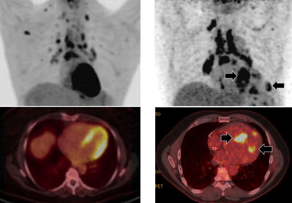
Image: The upper images shows PET scans, and the lower images combined PET/CT images of the heart. The images on the left show a patient on a 24-hour high-fat, low-sugar diet, while the right-hand images are those of a patient on a 72 hours high-fat, low sugar diet (Photo courtesy of UIC).
Researchers in the US have discovered that they can increase the certainty of diagnosing cardiac sarcoidosis, if from 72 hours until the diagnostic imaging exam the patients are subject to a high-fat, low-sugar diet.
Sarcoidosis is caused by the inflammation of a number of organs, including the lungs and lymph nodes, and can eventually affect the heart as well, and cause death from pulmonary fibrosis. Cardiac Sarcoidosis is most often diagnosed using Positron Emission Tomography (PET) and Computed Tomography (CT) scans, and a radioactive tracer called fludeoxyglucose (FDG), but the results are sometimes difficult to interpret.
The researchers from the University of Illinois at Chicago (UIC; Chicago, Illinois, USA) published the results of their research online in the December 2016 issue of the journal Clinical Nuclear Medicine.
To reduce the amount of sugar taken up by healthy heart cells during the PET imaging, clinicians often give patients a high-fat, low-sugar diet for 24 hours before the scan. Healthy cells take up fat, while diseased cells take up the tracer and sugar, and are more easily detected in the scans. The UIC team then tried to use a 72-hour high-fat, low-sugar diet, before the scans, in 215 tests from 207 patients, in the study period between 2014 and 2015. The results showed that 42% of patients with the 24-hour diet had indeterminate results, compared to only 4% of patients with a 72-hour diet. The 72-hour diet enabled the researchers to conclude with a high degree of confidence whether the patients had cardiac sarcoidosis or not.
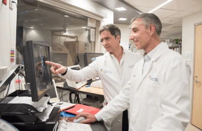
Corresponding author of the study, Dr. Yang Lu, assistant professor of radiology, UIC College of Medicine, said, "Up until now, diagnosis is made by expert opinion based on the results of several testing techniques including imaging. But what these scans show is often ambiguous, and so using them to definitively diagnose cardiac sarcoidosis is problematic. By following the diet for three days instead of one, the unpredictable but normal uptake of sugar by healthy heart cells was suppressed, so we could be more confident that areas that picked up the FDG had a very high likelihood of being affected by sarcoidosis, and this minimized the number of indeterminate findings where we can't make a diagnosis. The technique will also allow clinicians to better-follow their patients' progress under treatment for sarcoidosis."
Related Links:
University of Illinois at Chicago
Latest MRI News
- AI-Assisted Model Enhances MRI Heart Scans
- AI Model Outperforms Doctors at Identifying Patients Most At-Risk of Cardiac Arrest
- New MRI Technique Reveals Hidden Heart Issues
- Shorter MRI Exam Effectively Detects Cancer in Dense Breasts
- MRI to Replace Painful Spinal Tap for Faster MS Diagnosis
- MRI Scans Can Identify Cardiovascular Disease Ten Years in Advance
- Simple Brain Scan Diagnoses Parkinson's Disease Years Before It Becomes Untreatable
- Cutting-Edge MRI Technology to Revolutionize Diagnosis of Common Heart Problem
- New MRI Technique Reveals True Heart Age to Prevent Attacks and Strokes
- AI Tool Predicts Relapse of Pediatric Brain Cancer from Brain MRI Scans
- AI Tool Tracks Effectiveness of Multiple Sclerosis Treatments Using Brain MRI Scans
- Ultra-Powerful MRI Scans Enable Life-Changing Surgery in Treatment-Resistant Epileptic Patients
- AI-Powered MRI Technology Improves Parkinson’s Diagnoses
- Biparametric MRI Combined with AI Enhances Detection of Clinically Significant Prostate Cancer
- First-Of-Its-Kind AI-Driven Brain Imaging Platform to Better Guide Stroke Treatment Options
- New Model Improves Comparison of MRIs Taken at Different Institutions
Channels
Radiography
view channel
AI Hybrid Strategy Improves Mammogram Interpretation
Breast cancer screening programs rely heavily on radiologists interpreting mammograms, a process that is time-intensive and subject to errors. While artificial intelligence (AI) models have shown strong... Read more
AI Technology Predicts Personalized Five-Year Risk of Developing Breast Cancer
Breast cancer remains one of the most common cancers among women, with about one in eight receiving a diagnosis in their lifetime. Despite widespread use of mammography, about 34% of patients in the U.... Read moreMRI
view channel
AI-Assisted Model Enhances MRI Heart Scans
A cardiac MRI can reveal critical information about the heart’s function and any abnormalities, but traditional scans take 30 to 90 minutes and often suffer from poor image quality due to patient movement.... Read more
AI Model Outperforms Doctors at Identifying Patients Most At-Risk of Cardiac Arrest
Hypertrophic cardiomyopathy is one of the most common inherited heart conditions and a leading cause of sudden cardiac death in young individuals and athletes. While many patients live normal lives, some... Read moreUltrasound
view channel
Non-Invasive Ultrasound-Based Tool Accurately Detects Infant Meningitis
Meningitis, an inflammation of the membranes surrounding the brain and spinal cord, can be fatal in infants if not diagnosed and treated early. Even when treated, it may leave lasting damage, such as cognitive... Read more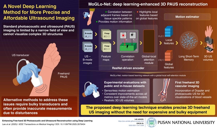
Breakthrough Deep Learning Model Enhances Handheld 3D Medical Imaging
Ultrasound imaging is a vital diagnostic technique used to visualize internal organs and tissues in real time and to guide procedures such as biopsies and injections. When paired with photoacoustic imaging... Read moreGeneral/Advanced Imaging
view channelAI Algorithm Accurately Predicts Pancreatic Cancer Metastasis Using Routine CT Images
In pancreatic cancer, detecting whether the disease has spread to other organs is critical for determining whether surgery is appropriate. If metastasis is present, surgery is not recommended, yet current diagnostic methods often miss these cases. As a result, many patients undergo unnecessary and invasive operations that... Read more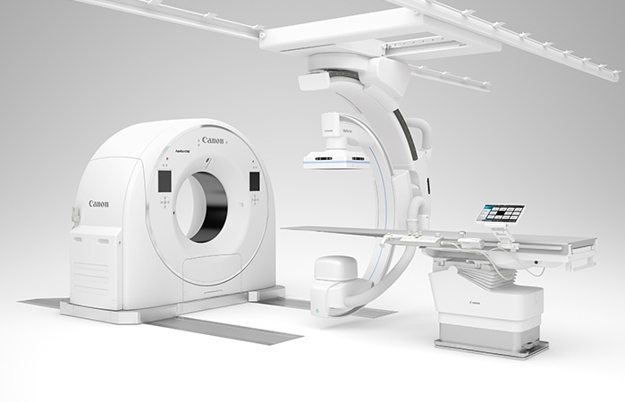
Cutting-Edge Angio-CT Solution Offers New Therapeutic Possibilities
Maintaining accuracy and safety in interventional radiology is a constant challenge, especially as complex procedures require both high precision and efficiency. Traditional setups often involve multiple... Read moreImaging IT
view channel
New Google Cloud Medical Imaging Suite Makes Imaging Healthcare Data More Accessible
Medical imaging is a critical tool used to diagnose patients, and there are billions of medical images scanned globally each year. Imaging data accounts for about 90% of all healthcare data1 and, until... Read more
Global AI in Medical Diagnostics Market to Be Driven by Demand for Image Recognition in Radiology
The global artificial intelligence (AI) in medical diagnostics market is expanding with early disease detection being one of its key applications and image recognition becoming a compelling consumer proposition... Read moreIndustry News
view channel
GE HealthCare and NVIDIA Collaboration to Reimagine Diagnostic Imaging
GE HealthCare (Chicago, IL, USA) has entered into a collaboration with NVIDIA (Santa Clara, CA, USA), expanding the existing relationship between the two companies to focus on pioneering innovation in... Read more
Patient-Specific 3D-Printed Phantoms Transform CT Imaging
New research has highlighted how anatomically precise, patient-specific 3D-printed phantoms are proving to be scalable, cost-effective, and efficient tools in the development of new CT scan algorithms... Read more
Siemens and Sectra Collaborate on Enhancing Radiology Workflows
Siemens Healthineers (Forchheim, Germany) and Sectra (Linköping, Sweden) have entered into a collaboration aimed at enhancing radiologists' diagnostic capabilities and, in turn, improving patient care... Read more


