Flexible Ultrasound Patch Monitors Cardiovascular Function
By MedImaging International staff writers
Posted on 12 Aug 2021
A novel wearable skin patch that incorporates an array of ultrasonic transducers can provide continuous monitoring of deep-tissue hemodynamics, claims a new study.Posted on 12 Aug 2021
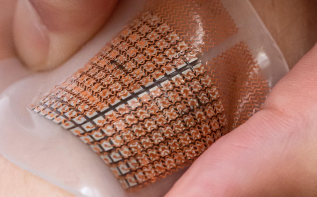
Developed at the University of California, San Diego (UCSD; USA), Yonsei University (Seoul, South Korea), the Korea Institute of Science and Technology (KIST; Seoul, Republic of Korea), and other institutions, the patch is made of a flexible, stretchable polymer that that is embedded with a 12X12 grid of millimeter-sized ultrasound transducers, in a phased array design. When electricity flows through the transducers, they emit ultrasound waves that travel through the skin and deep into the body.

The phased array ultrasound transducer patch (Photo courtesy of UCSD)
The computerized phased array design has two main modes of operation. In one, the transducers are synchronized to transmit ultrasound waves together, producing a high-intensity ultrasound beam that focuses on one spot up to 14 centimeters in the body. In the other mode, the transducers can be programmed to transmit out of sync, allowing for active focusing and steering of ultrasound beams over a range of incident angles so as to target various regions of interest. When the waves penetrate through a major blood vessel, they encounter movement from red blood cells (RBCs) flowing inside.
These movement changes or shifts, known as Doppler frequency shift, reflect back to the patch, and are used to create a visual recording of the blood flow. This same mechanism can also be used to create moving images of the heart’s walls. In a study conducted in healthy volunteers, the phased array patch monitored Doppler spectra from cardiac tissues, recorded central blood flow waveforms, and estimated cerebral blood supply in real time. The study was published on July 16, 2021, in Nature Biomedical Engineering.
“Sensing signals at such depths is extremely challenging for wearable electronics. Yet, this is where the body’s most critical signals and the central organs are buried,” said co-first author nanoengineer Chonghe Wang, PhD, of UCSD. “We engineered a wearable device that can penetrate such deep tissue depths and sense those vital signals far beneath the skin. This technology can provide new insights for the field of healthcare.”
Related Links:
University of California
Yonsei University
Korea Institute of Science and Technology


