Shoulder Brightness on Ultrasound Could Indicate Diabetes
By MedImaging International staff writers
Posted on 04 Dec 2018
Unusually bright (hyperechoic) ultrasound images of the deltoid muscle may be a warning sign of type 2 diabetes (T2D), according to a new study.Posted on 04 Dec 2018
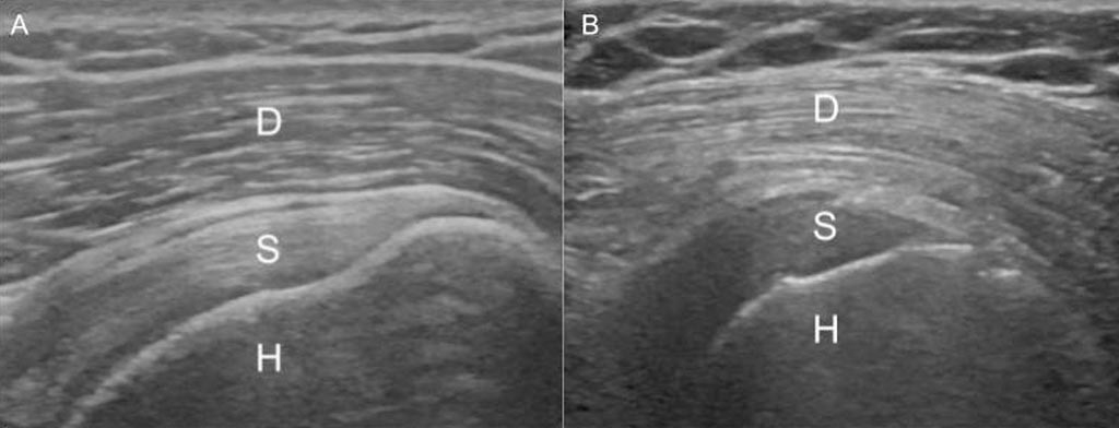
Researchers at Henry Ford Hospital (Detroit, MI, USA) compiled 137 shoulder ultrasounds from patients with T2D, including 13 with pre-diabetes; they also obtained 49 ultrasounds from obese patients without diabetes. The ultrasounds were shown to two musculoskeletal radiologists who were asked to classify the patients, based on the brightness of their shoulder muscle, into one of three categories: normal, suspected diabetes, and definite diabetes. A third musculoskeletal radiologist acted as an arbitrator when the other two disagreed.

Image: A displays the normal gradient of the deltoid muscle; B shows reversal in T2D patient. D: Deltoid, S: Supraspinatus, H: Humerus (Photo courtesy of RSNA).
Based on the shoulder ultrasounds, the radiologists correctly predicted diabetes in 70 of 79 patients (89%). Brightness on ultrasound was also an accurate predictor of pre-diabetes, with all 13 pre-diabetic patients assigned to either suspected or definite diabetes categories. The researchers suspect that the hyperechoic image is due to low or depleted levels of glycogen in the muscle, which are decreased in T2D patients by up to 65%. The study was presented at the annual meeting of the Radiological Society of North America (RSNA), held during November 2018 in Chicago (IL, USA).
“We weren't surprised that we had positive results, because the shoulder muscle on patients with diabetes looked so bright on ultrasound, but we were surprised at the level of accuracy. A lot of the patients weren't even aware that they were diabetic or pre-diabetic,” said senior author and study presenter musculoskeletal radiologist Steven Soliman, DO. “If we observe this in patients with pre-diabetes and diabetes, we can get them to exercise, make diet modifications and lose weight. If these interventions happen early enough, the patients may be able to avoid going on medications and dealing with all the complications that go with the disease.”
Glycogen is a multi-branched polysaccharide that forms the main storage form of glucose in the body. In humans, glycogen is made and stored primarily in the cells of the liver and skeletal muscle. Muscle cell glycogen appears to function as an immediate reserve source of available glucose for muscle cells. As muscle cells lack glucose-6-phosphatase, which is required to pass glucose into the blood, the glycogen they store is available solely for internal use and is not shared with other cells.
Related Links:
Henry Ford Hospital


