Contrast-Enhanced Ultrasound Helps Detects Liver Cancer
By MedImaging International staff writers
Posted on 14 Nov 2017
A new study reveals that contrast-enhanced ultrasound (CEUS) can achieve correct diagnosis without radiation, and at lower cost.Posted on 14 Nov 2017
Researchers at the University of Calgary (Alberta, Canada) and Stanford University (CA, USA) conducted a study in 200 patients at risk for hepatocellular carcinoma in order to assess detection and characterization of malignant focal liver lesions using CEUS, an imaging technique that allows pathology detection without radiation, expensive magnetic resonance imaging (MRI) equipment, or biopsies. The researchers also utilized the Liver Imaging Reporting and Data System (LI-RADS), a tool used to classify liver tumors using computed tomography (CT), MRI, and CEUS.

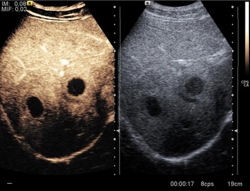
Image: A new study suggests contrast enhance ultrasound (L) provides good diagnostic aid (Photo courtesy of radiopaedia.org).
The results revealed that CEUS can provide dynamic real-time imaging with high spatial and temporal capability, and with 97% accuracy. The researchers suggest that in liver cancer patients with lesions that are indeterminate on CT and MRI, CEUS can serve as a useful tool to improve management and assist guidance of liver biopsies and local treatment of hepatocellular cancer. The study was presented at the 32nd annual conference of the International Contrast Ultrasound Society (ICUS), held during October 2017 in Chicago (IL, USA).
“Patients with hepatocellular carcinoma, the third leading cause of cancer deaths worldwide, were found to benefit from contrast-enhanced ultrasound imaging when magnetic resonance imaging was inconclusive,” said senior author Professor Stephanie Wilson, MD, of the University of Calgary, and co-president of ICUS. “This is an exciting option, because hepatocellular carcinoma is the most common form of liver cancer, and standard imaging with MRI is often an insufficient option for characterizing the tumor.”
CEUS uses liquid suspensions of miniscule gas microbubbles to improve the clarity and reliability of an ultrasound image. The microbubbles are smaller than red blood cells, and when they are injected into a patient's vein, they flow through the microcirculation and reflect ultrasound signals, thus improving the accuracy of diagnostic ultrasound exams. The microbubbles are expelled from the body within minutes.
Related Links:
University of Calgary
Stanford University


 Guided Devices.jpg)


