Hybrid Imaging Technology Developed for Improved Identification of Atherosclerotic Plaques
By MedImaging International staff writers
Posted on 01 Jun 2016
Collaborative research between two US NIBIB-funded laboratories has resulted in a new method for identifying atherosclerotic plaques.Posted on 01 Jun 2016
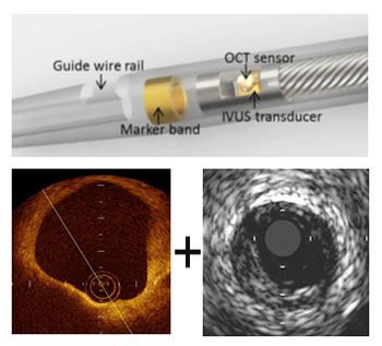
The technique uses two different types of imaging, and provides significantly more depth and detail than existing methods. The technique provides clinicians with an improved diagnostic tool for finding problematic plaques, for example the Thin-Cap Fibroatheroma (TCFA).

Image: The IVUS-OCT device can capture infrared and ultrasound images, and help find plaque that could cause a heart attack or stroke (Photo courtesy of the US National Institute of Biomedical Imaging and Bioengineering).
The hybrid imaging technology called IntraVascular Ultrasound and Optical Coherence Tomography (IVUS-OCT) is licensed by OCT Medical Imaging (Irvine, CA, USA), and consists of an ultrafast optical-ultrasonic system, and a miniaturized catheter that can be used to image and characterize atherosclerotic plaques in vivo.
The research by the US National Institute of Biomedical Imaging and Bioengineering (NIBIB; Bethesda, MD, USA) laboratories was published in the December 18, 2015, issue of the journal Scientific Reports.
Zhongping Chen, PhD, senior author of the paper, said, “This work has overcome many of the challenges of doing IVUS and OCT simultaneously. When you use a single catheter, you don’t have to worry about matching two images, which enables physicians to look at co-registered ultrasound and OCT images simultaneously in real time. It will make identifying plaques easier and more precise, allowing for better treatment decisions.”
Related Links:
OCT Medical Imaging
US National Institute of Biomedical Imaging and Bioengineering


