Ultrahigh-Frequency Fine Tunes Ultrasound Imaging
By MedImaging International staff writers
Posted on 12 May 2016
An innovative ultrahigh-frequency (UHF) ultrasound system offers exceptional detail with resolution as fine as 30 micrometers, the equivalent of less than half the size of a grain of sand.Posted on 12 May 2016
The Fujifilm VisualSonics (Toronto, ON, Canada) Vevo MD system is designed to play a role in a range of specializations, including neonatology, vascular, musculoskeletal, dermatology, and other clinical applications that pertain to the first three cm of the body. The system is capable of operating in a range of UHF frequencies of up to 70 MHz, when used with the Fujifilm VisualSonics UHF series of transducers.

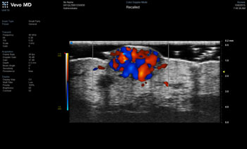
Image: A superficial hemangioma with color created with the Vevo MD (Photo courtesy of Fujifilm VisualSonics).
“As the recognized leader in ultra high frequency imaging systems, Fujifilm VisualSonics is proud to be the first to market with unparalleled technology,” said Renaud Maloberti, vice president and general manager of Fujifilm VisualSonics. “With the Vevo MD, clinicians can observe the tiniest, most highly detailed anatomy that has never been seen before, which means greatly enhanced potential for diagnoses.”
“The Vevo high frequency ultrasound systems and linear array transducers are ideal for fast, high resolution imaging,” said assistant professor Craig Goergen, PhD, of Purdue University (Lafayette, IN, USA). “The ease of use and unmatched image quality allow for visualization of fine structures, opening up numerous biomedical applications for us to explore.”
While the UHF ultrasound frequency (100 MHz – 1 GHz) has been used in non-destructive evaluation of materials and biomedical imaging for many years, its applications in biology and medicine have so far been limited. With the advent of UHF ultrasound, the width of the ultrasound beam is of only a few microns, approaching the dimensions of many cells, which allows the detection of structure too small for conventional ultrasound, such as pseudoaneurysms in artery walls.
Related Links:
Fujifilm VisualSonics


 Guided Devices.jpg)


