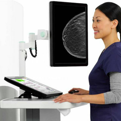
AI-Based Algorithm Improves Accuracy of Breast Cancer Diagnoses
Posted on 26 Aug 2024
High breast tissue density is known to raise the risk of breast cancer, and its measurement can be derived from mammograms. Accurate interpretation of mammograms is vital for successful breast cancer screening but is hampered by inconsistencies in radiological evaluations and a worldwide shortage of radiologists. To overcome these challenges, a new artificial intelligence (AI)-based algorithm uses deep learning to analyze multiple mammogram views concurrently, simulating the evaluation process of radiologists.
The MV-DEFEAT algorithm, developed by researchers at the University of Eastern Finland (Kuopio, Finland), improves the assessment of mammogram density, potentially revolutionizing radiological procedures by facilitating more accurate diagnoses. The research team applied a multi-view deep evidential fusion approach, incorporating aspects of the Dempster-Shafer evidential theory and subjective logic to provide a thorough analysis of mammogram images from various angles. MV-DEFEAT significantly outperforms existing methods, increasing the precision of mammogram screenings by effectively and consistently determining the density and distribution of dense breast tissue. Specifically, in the public VinDr-Mammo dataset, which contains over 10,000 mammograms, the algorithm achieved a 50.78% improvement in differentiating between benign and malignant tumors compared to previous multi-view approaches. Additionally, MV-DEFEAT generates gradient-based saliency maps that accentuate critical areas, aiding radiologists in their diagnostic decisions.
")
The algorithm's effectiveness across various datasets highlights its ability to accommodate diverse patient demographics. It was tested using data from four open-source datasets, which broadened its accuracy and generalizability. These features underscore the potential of AI-driven methods in medical diagnostics. Although MV-DEFEAT significantly supports breast cancer screening, the researchers stress the need for ongoing enhancement and validation to confirm its dependability and effectiveness in clinical environments. This progress represents a critical step toward integrating AI in diagnostic procedures, which may lead to earlier detection and improved outcomes for breast cancer patients.
“To fully integrate AI like MV-DEFEAT into clinical practice, it is crucial to build trust among healthcare professionals through rigorous testing and validation,” said Doctoral Researcher Raju Gudhe of the University of Eastern Finland. “Indeed, our next steps involve further validation studies to establish MV-DEFEAT as a reliable tool for breast cancer diagnostics in Finland.”
Related Links:
University of Eastern Finland