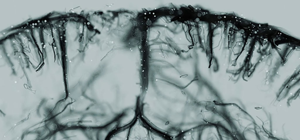
Major Advance in 3D Ultrasound Imaging Enables Real Time Observation of Blood Flow
Posted on 16 May 2022
Organs are supplied by a complex network of blood vessels, which are essential for their proper functioning. Some imaging techniques give a global view of this vascular network, but for the first time, ultrafast 3D imaging allows us to observe blood flow from the large arteries to the smallest blood vessels of only a few micrometers in diameter.
Two successive studies by researchers at the Physics for Medicine Paris (ESPI, Paris, France) have highlighted advances in non-invasive 3D ultrasound imaging, making it possible to observe blood flow in real time in two whole organs: the heart and the brain. Over the last 10 years, ESPCI has made major advances in vascular imaging, with the development of ultrasensitive Doppler imaging (uDoppler) and then ultrasound localization microscopy (ULM) in 2D. This time, the researchers at ESPCI have reached a major milestone by deploying ULM in 3D: thanks to the three-dimensional aspect, the researchers obtained super-resolved images of the rodent heart and brain, at the scale of the entire organ. In addition to providing fundamental knowledge of organ function, this technique could also provide valuable information on various cardiovascular pathologies and even measure the effectiveness of different treatments.

To achieve such a feat at such fine resolutions, the scientists injected microscopic gas bubbles, the position of which was monitored at high imaging rates. This made it possible to obtain detailed information on blood flow and channel size, and thus to reconstruct the entire vascular activity of the organ. The team also had to overcome several technological challenges. For the heart, for example, it was necessary to find the ideal measurement window to be able to correct the movements linked to breathing and heartbeats on the image. For the brain, it may be necessary to implement post-processing algorithms to correct signal distortions induced by the skull. Moreover, the transition from 2D to 3D imaging implies a huge increase in the volume of data collected: for one minute of acquisition, the volume of data to be processed exceeds one terabyte of information. Before considering a move into the human clinic, the scientists will further improve their technology by optimizing the sensors, electronics and data processing methods.
Related Links:
ESPI


