In-Office Solution Provides Direct Arthroscopic Imaging
By MedImaging International staff writers
Posted on 27 Mar 2018
A novel handheld tool provides illumination and visualization of interior cavities of the body through either a natural or surgical opening.Posted on 27 Mar 2018
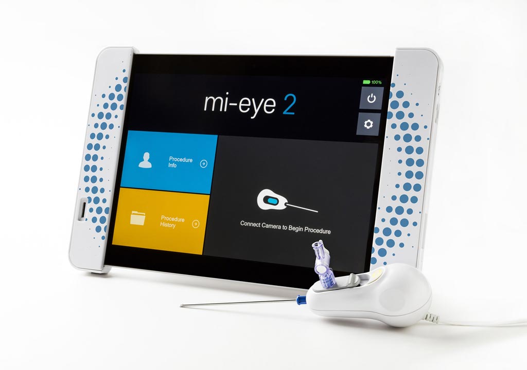
The Trice Medical (Malvern, PA, USA) Mi-Eye 2 is a single-use, disposable arthroscope that consists of a needle with an integrated camera and light source that allows surgeons to perform a full diagnostic arthroscopic or endoscopic procedure as an alternative to traditional diagnostic modalities, such as time-consuming magnetic resonance imaging (MRI). Features of the Mi-Eye 2 device include a 120° field of view, a 5-35mm depth of field, real-time image and video capture, and integrated optic and light source. The images are viewed on a dedicated high definition (HD) tablet device with a 10.8” display powered by a 1.6GHz Intel atom processor.

Image: The Mi-Eye 2 handheld visualization device and dedicated HD tablet (Photo courtesy of Trice Medical).
The heart of the system is a retractable 2.2 mm needle percutaneously inserted into the joint space, with the joint numbed prior to the procedure using local anesthesia. Once inside the joint space, the needle retracts to expose the Mi-Eye 2 camera and light source; once retracted, the needle is blunted by the camera. The surgeon can then perform a diagnostic arthroscopy exam to detect pathology in the intra-articular space. A 19-gauge lumen is available to inject or aspirate fluid during the procedure. At the conclusion of the procedure, the entirety of the Mi-Eye 2 is thrown into a sharps container.
“MRI and Ultrasound will always be good options for patients with sports injuries. We are thrilled to add disposable in-clinic arthroscopy to the list of available tools physicians have to be able to assess joint injuries,” said Mark Foster, chief commercialization officer and VP of worldwide sales at Trice Medical. “In many countries, the Mi-Eye 2 has a chance to save weeks to months of the treatment pathway to surgery and provide a dynamic image of the joint.”
“The Mi-Eye 2 offers results better than those of traditional MRI scans for diagnosing certain conditions; it provides an eye inside the body. The advantage of the technology is that it takes places in a setting convenient for the patient. They find out their results right on the spot,” said orthopedic surgeon John Corsetti, MD, of New England Orthopedic Surgeons (NEOS; Springfield, MA, USA). “Patient satisfaction has been extremely high with this new technology. They are pleased that they get to work with me while they are awake so they can ask me any questions about what they see on the screen.”
Related Links:
Trice Medical


