MRI

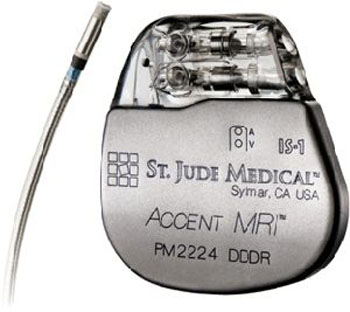
First Implantable Cardioverter-Defibrillator System to Allow for Full-Body MRI Scans Launched
A new implantable cardioverter-defibrillator system is the first to combine effective treatment performance, improved comfort, increased longevity, with full-body magnetic resonance imaging access. More...01 May 2014


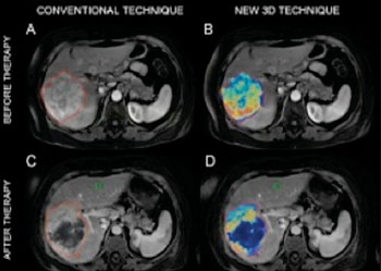
3D MRI Offers Improved Prediction of Survival After Chemotherapy for Liver Tumors
Researchers are using specialized 3D MRI scanning technology to accurately measure living and dying liver tumor tissue in order to quickly show whether very toxic chemotherapy, delivered right through a tumor’s blood supply, is working. More...17 Apr 2014
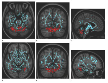
MRI’s Magnetic Fields Used to Treat Balance Problems
Investigators, expanding on earlier studies, reported that individuals with balance disorders or dizziness traceable to an inner-ear disturbance show distinctive abnormal eye movements when the affected ear is exposed to the strong pull of a magnetic resonance imaging scanner’s magnetic field. More...15 Apr 2014
In Other News
MR Array Designed for Imaging the Orbit and Mandible
CE Marking Approval and First Implants of MRI Pacing Leads Announced
7-Tesla MRI Technology Offers More Precise Window into the Brain
MRI Identifies Parkinson’s in Patients at Risk of Dementia
Ultrahigh-Field MRI Shows Potential for Earlier Diagnosis of Parkinson’s Disease
Magnetoencephalography/MRI Combo Offers Real-time Clues into Brain Functioning
Ultrahigh Field MRI Technology Provides More Effective Diagnosis of Breast Cancer
First 4-F Cardiac Resynchronization MR Conditional Lead Receives CE Approval
Carotid Artery MRI May Predict Probability of Heart Attack, Stroke
Hyperpolarization MRI Scanning Technology Can Visualize Metabolic Changes
fMRI Findings Reveal Impact of Head Movement in Multiple Sclerosis Patients
MR Technology Provides Radiation-Free Imaging of Childhood Tumors
First MR-Safe and CT-Compatible Neurosurgical Horseshoe Headrest Provides Nonrigid Positioning
Market Agreement Established for Next-Generation Software to Enable MRI-Guided, Catheter-Based Procedures
MRI Study to Offer New Strategies for Chronic Kidney Disease Treatments
4D Blood Flow MRI Technology Detects Aortic Disease and May Predict Complications

MR Radiation Oncology Suite Provides Consistent Patient Positioning, Effectively Characterizes Cancer
Multifunctional Nanoparticle Targets Blood Vessel Plaques
First Lung Cancer Patients Treated Using MRI-Guided Radiotherapy
Imaging Shows Unique Brain Region Tied to Higher Cognitive Abilities
MRI and Automated Surface Mesh Modeling Analysis Reveal Changes in Brain Anatomy in Women with Multiple Sclerosis and Depression
Imaging Technologies That Predict Cognitive Development Studied
Agreement to Expand Distribution for MR Volumetric Brain Atrophy Software
MedImaging's MRI channel in addition to reporting on MR hardware, informs about the many magnetic resonance applications possible with the technique notably MRI, fMRI, diffusion MRI, MR angiography, MR guided surgery, in addition to industry developments, and safety issues.


