New Ultrasound Technologies Improve Diagnosis for Cancer, Liver Disease and Other Pathologies
Posted on 17 Sep 2024
Several diseases, including some cancers, can remain hidden or difficult to detect using traditional medical imaging. However, new technologies developed by researchers may soon enhance ultrasound's effectiveness in diagnosing conditions such as cancer, liver disease, and other pathologies.
The United States Patent and Trademark Office has recently granted four patents for advanced diagnostic ultrasound technology created by researchers at the University of Rochester (Rochester, NY, USA). Using advanced physics, mathematics, and scattering theory, the researchers have developed techniques to extract previously hidden features from ultrasound data, revealing potential issues with organs like the liver, thyroid, or breast. Some of these innovations have already been licensed to startups aiming to bring these advancements into clinical settings to benefit patients.

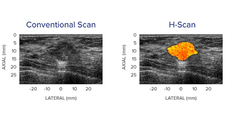
Two of the patents are associated with the H-scan technique, while the other two focus on reverberant shear wave fields. The H-scan technology enhances traditional black-and-white ultrasound images by adding color to specific features—for example, coding fat deposits in the liver as yellow or indicating cancer in the breast with red. The technologies related to reverberant shear wave fields improve elastography, which measures tissue stiffness. These advancements provide quicker, more cost-effective methods for delivering critical diagnostic information to doctors and radiologists. Since these technologies focus on ultrasound image processing, they can be seamlessly integrated into existing ultrasound equipment without the need for new hardware.
“These are inventions that you can retrofit to existing imaging systems. You can reprogram the scanners to process our way and out comes this new analysis and information,” said Kevin Parker, the William F. May Professor of Engineering at the University’s Hajim School of Engineering & Applied Sciences, who has developed the ultrasound technologies. “We don’t have to recreate a whole new generation of ultrasound scanners.”
Related Links:
University of Rochester


