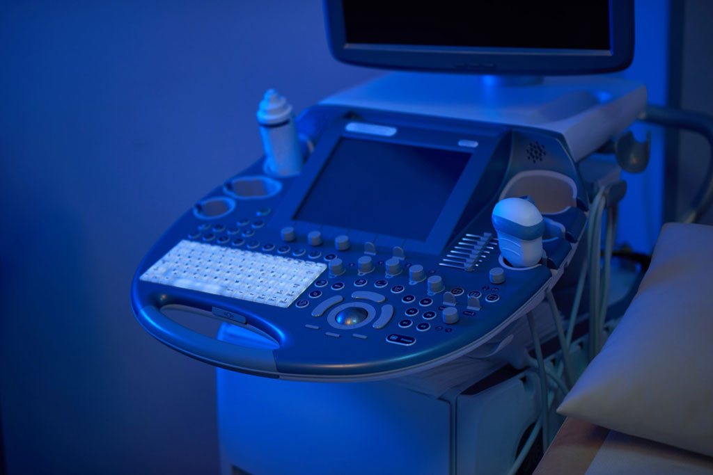
AI Beats Sonographers in Assessing Heart Health from Echocardiogram Images
Posted on 10 Apr 2023
The first symptoms of heart trouble can often be severe illness or even death. While doctors and scientists have persistently improved models and techniques for predicting heart disease risk, artificial intelligence (AI) has emerged as a powerful ally. The debate over whether AI or a sonographer is better at evaluating and diagnosing cardiac function after analyzing an echocardiogram has remained unresolved. Now, a new study has demonstrated that AI is superior to sonographers in assessing and diagnosing cardiac function through echocardiograms.
The first-of-its-kind, blinded, randomized clinical trial of AI in cardiology was led by investigators at Cedars-Sinai (Los Angeles, CA, USA) who are optimistic that the technology will prove advantageous when implemented throughout the Cedars-Sinai clinical system and health systems nationwide. In 2020, the team developed one of the first AI technologies for evaluating cardiac function, specifically focusing on left ventricular ejection fraction, a crucial measurement used in cardiac function diagnosis. Expanding on these findings, the new study examined the accuracy of AI in assessing 3,495 transthoracic echocardiograms by comparing initial evaluations made by AI or a sonographer (an ultrasound technician).

The study found that cardiologists more often agreed with AI's initial assessments, making corrections to only 16.8% of AI-generated evaluations compared to 27.2% of those made by sonographers. The study also found that physicians could not distinguish between assessments made by AI and those made by sonographers. Furthermore, AI assistance saved time for both cardiologists and sonographers. The researchers hope that AI support can help clinicians save time and reduce the more monotonous aspects of the cardiac imaging workflow. However, they maintain that the cardiologist should remain the ultimate expert in evaluating AI model output.
“The results have immediate implications for patients undergoing cardiac function imaging as well as broader implications for the field of cardiac imaging,” said cardiologist David Ouyang, MD, principal investigator of the clinical trial and senior author of the study. “This trial offers rigorous evidence that utilizing AI in this novel way can improve the quality and effectiveness of echocardiogram imaging for many patients.”
Related Links:
Cedars-Sinai


