AI Tool Simplifies Thyroid Ultrasound Scanning Workflow
By MedImaging International staff writers
Posted on 17 May 2021
A new artificial intelligence (AI) algorithm views and quantifies ultrasound image data to aid rapid diagnosis of thyroid gland nodules.Posted on 17 May 2021
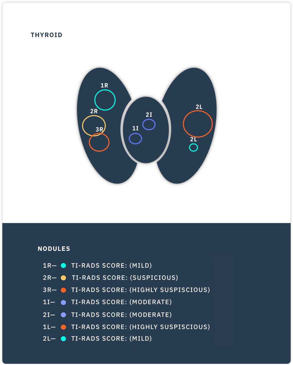
The Medo (Edmonton, Canada) Medo-Thyroid software solution uses AI and machine learning to radically simplify the classification of thyroid nodules. Users simply save sweeps of each thyroid lobe, and Medo automatically analyses these to select the most relevant image of the nodule and calculate standard lobe and isthmus measurements. It also segments the nodule (across all images in the sweep) in order to calculate actual volumetric data, as required for TI-RADS scoring, and finally to create individualized interactive reports.

Image: An example of a Medo-Thyroid interactive report (Photo courtesy of Medo)
“Medo Thyroid makes thyroid ultrasound a less frustrating test, by presenting thyroid measurements and nodule characteristics in a convenient format. It's especially helpful to radiologists when following up multiple nodules,” said radiologist Jacob Jaremko, MD, co-founder of Medo. “Medo-Thyroid contains several key technological breakthroughs, including a cross-referencing ability previously only possible on multi-plane CT and MRI. This feature is designed to assist the user with viewing individual nodules across all planes of interest, such as transverse and sagittal.”
“Using artificial intelligence, Medo has revolutionized the process of performing thyroid studies and turned it into a seamless, fast, and objective workflow,” said Dornoosh Zonoobi, MD, co-founder and CEO of Medo. “We are particularly pleased with the feedback we have received from many of the independent clinicians who have used our product, stating that this will greatly benefit them in their everyday workflow.”
Over 1.5 million thyroid ultrasound exams are performed each year in the United States alone. The high volume is due to the fact that nodules on the thyroid gland are common, and although usually benign, they can be cancerous. Ultrasound exam of the thyroid is time consuming and potentially error-prone, and usually involves tedious review of hundreds of images in order to locate and measure each nodule individually. The process becomes even more difficult--and subjective--when the gland has multiple nodules.
Related Links:
Medo


