Ultrasound Bone Assessment Could Increase Osteoporosis Screening
By MedImaging International staff writers
Posted on 06 Mar 2019
A new study suggests that calcaneus ultrasonography (US) can establish bone mineral density (BMD) on par with gold standard dual-energy x-ray absorptiometry (DXA).Posted on 06 Mar 2019
Researchers at the Louis Stokes Cleveland VA Medical Center (OH, USA), the West Virginia School of Osteopathic Medicine (WVSOM; Lewisburg, USA), and other institutions conducted a study involving 99 patients at a rural primary care facility in order to establish data ranges from US of the calcaneus (heel bone) that correspond to BMD stratification as identified by DXA, and to determine whether vitamin D concentration adds to US bone health assessment.

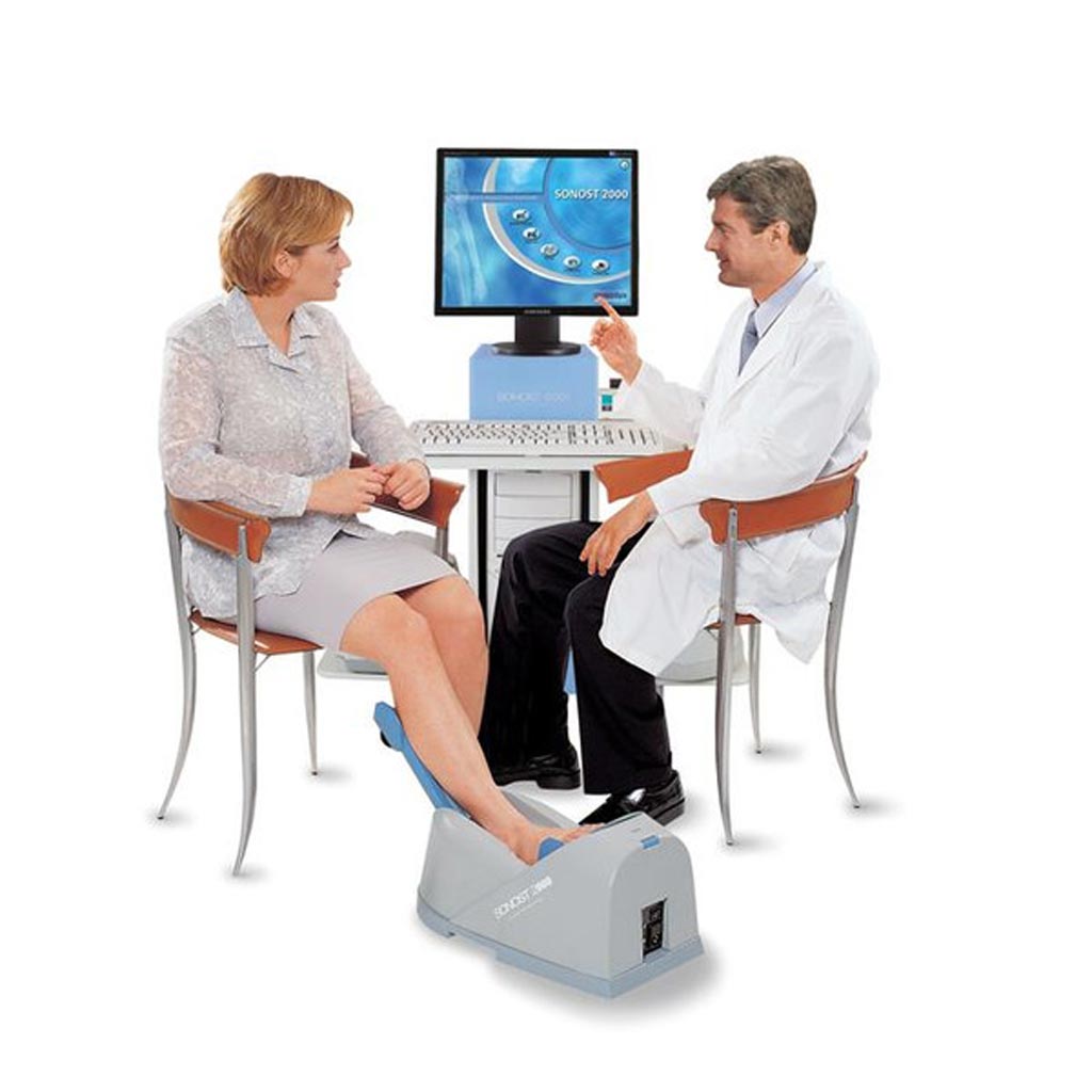
Image: The Sonost 2000 calcaneus ultrasound bone densitometer (Photo courtesy of Econet).
Ultrasonography was used to scan the left and right calcaneus, and blood was collected for vitamin D analysis. Other data collected included fracture risk assessment tool parameters, menstrual history, and drug and supplement use. The researchers then calculated correlations within and between DXA and US measurements, as well as correlations between DXA, US measurements, and vitamin D levels. Finally, predictive performance of US readings on bone health (as determined by DXA scan) was assessed.
The results revealed that US readings of either the left or right foot were predictive of bone quality, with no differences found between them. There was no correlation found between DXA- and US-assessed BMD and vitamin D concentrations. The researchers added that while DXA scans remain the best option for thorough, comprehensive information on BMD, the equipment is expensive, immobile, and exposes patients to ionizing radiation, creating barriers to screening larger populations. The study was published in the March 2019 issue of the Journal of the American Osteopathic Association.
“Using ultrasound to scan the heel won't give us all the information we could gather with a full DXA scan. However, it gives us a clear enough snapshot to know whether we should be concerned for the patient,” concluded lead author associate professor of biomedical sciences Carolyn Komar, PhD, of WVSOM, and colleagues. “The affordability and mobility of a US machine enables its use as a screening method that may be applicable to large numbers of people.”
Unlike DXA, US does not measure BMD directly, but can provide indirect information on BMD, as well as more direct information on bone microarchitecture, such as trabecular number, connectivity, and orientation. It is important to remember that though indicative, an increase in BMD may not necessarily result in an improvement in bone strength, as the improved area may be the discontinuous end of a bony trabeculum, as opposed to strengthening a joined trabecular structure.
Related Links:
Louis Stokes Cleveland VA Medical Center
West Virginia School of Osteopathic Medicine


