Advanced Technology Maps Complex Cardiac Arrhythmias
By MedImaging International staff writers
Posted on 06 Nov 2017
An innovative high-resolution ultrasound-based electrophysiology system visualizes cardiac anatomy and maps dipole density to chart the pathway of every heartbeat.Posted on 06 Nov 2017
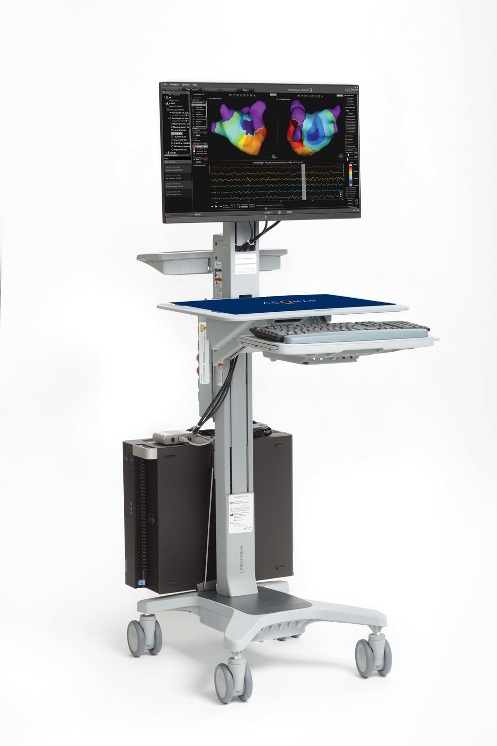
The Acutus Medical (Carlsbad, CA, USA) AcQMap High Resolution Imaging and Mapping System detects and displays both standard voltage-based and higher resolution dipole density (charge-source) maps of the human heart. The system combines ultrasound anatomy construction with an ability to map the electrical-conduction of each heartbeat, in order to identify complex arrhythmias across the entire atrial chamber. Following each ablation treatment, the heart can be re-mapped in real-time in just a few seconds to continually visualize any changes from the prior mapping.

Image: The AcQMap high-resolution imaging and mapping system workstation (Photo courtesy of Acutus Medical).
Using ultrasound, electrophysiologists can acquire more than 115,000 points every minute, providing computerized tomography (CT)-quality cardiac chamber reconstructions with electrical activity displayed as waveform traces. Dynamic, three-dimensional (3D) dipole density maps are overlaid on the cardiac chamber reconstruction to show chamber-wide electrical activation. The system can be used with all existing commercially available cardiac ablation platforms, and allows remapping of the entire chamber at any time during the procedure.
“The AcQMap System was designed in close collaboration with some of the most respected names in the field to provide practitioners with a suite of tools that enables them to rapidly map and re-map to visualize changes throughout the ablation procedure,” said Steven McQuillan, senior VP of regulatory and clinical affairs at Acutus Medical. “We firmly believe that by working together with EP practitioners and scientists, we will continue to uncover breakthrough innovations to improve and advance cardiac care.”
“The AcQMap System is able to provide global dipole density mapping of irregular and chaotic activation in the atrial chambers, whereas conventional sequential mapping may struggle to provide us with the information that is required,” said Tom Wong, MD, of Royal Brompton Hospital (London, United Kingdom). “In the cases we have performed thus far, real-time mapping of complex arrhythmias has allowed us to focus on areas of interest and terminate the arrhythmia using ablation therapy. We can now offer individualized, tailored therapy, and are one step closer to identifying the mechanisms of complex arrhythmias.”
Cardiac mapping collects and displays electroanatomical maps of the heart, and includes activation, isochronal, propagation, or voltage maps. Isochronal vector maps are commonly used to study the mechanisms and to guide the ablative therapies of arrhythmias; activation maps display local activation time, color-coded and overlaid on reconstructed 3D geometry; propagation maps show a dynamic color display of the propagation of the activation wavefront across the reconstructed chamber; and voltage map displays the peak-to-peak amplitude at each site.
Related Links:
Acutus Medical


