Portable Ultrasound Device Scans Neonatal Brain Activity
By MedImaging International staff writers
Posted on 26 Oct 2017
A novel device uses functional ultrasound imaging (fUSI) and video electroencephalography (EEG) to monitor brain microvasculature in newborns.Posted on 26 Oct 2017
Researchers at Paris Sciences et Lettres Research University (PSL; Paris, France), the University of Geneva (Switzerland), and other institutions used a lightweight (40 grams), flexible, and noninvasive headmount in order to simultaneously perform continuous video EEG and fUSI ultrafast Doppler (UfD) imaging of the brain microvasculature in human neonates. The system was able to detect very small cerebral blood volume variations that closely correlated with two different sleep states, as defined by EEG recordings.

Image: New research shows fusing ultrasound and EEG can help map seizures (Photo courtesy of Charlie Demene/ PSL).
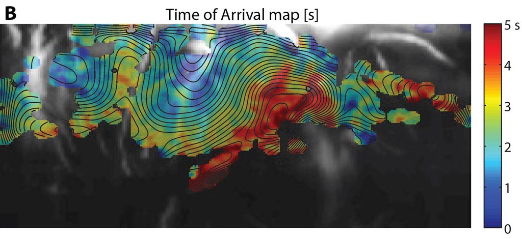
Using fUSI, the researchers were able to assess brain activity in two neonates with congenital abnormal cortical development, establishing neonatal seizure dynamics with high spatiotemporal resolution (200 μm for UfD and 1 ms for EEG). fUSI was then applied to track how waves of vascular changes were propagated during interictal periods, and to determine the ictal foci of the seizures. The new technology may potentially pave the way to therapeutic innovations for neurological disorders. The study was published on October 11, 2017, in Science Translational Medicine.
“Functional neuroimaging modalities are crucial for understanding brain function, but their clinical use is challenging,” concluded lead author Charlie Demene, PhD, of the PSL École Supérieure de Physique et de Chimie Industrielles (ESPCI), and colleagues. “Imaging the human brain with fUSI enables high-resolution identification of brain activation through neurovascular coupling, and may provide new insights into seizure analysis and the monitoring of brain function.”
Related Links:
Paris Sciences et Lettres Research University
University of Geneva


