Results of PRI-MUS High Resolution Micro-Ultrasound Protocol Published
By MedImaging International staff writers
Posted on 02 Aug 2016
Initial results supporting the development of a Prostate Risk Identification using Micro-Ultrasound (PRI-MUS) protocol are expected to result in a new standard for micro-ultrasound targeted prostate biopsies.Posted on 02 Aug 2016
The initial results supporting the development of the PRI-MUS protocol were published in a peer-reviewed article in the July 2016 issue of the journal Urology.

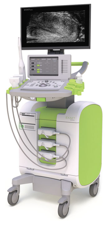
Image: The ExactVu micro-ultrasound imaging system (Photo courtesy of Exact Imaging).
The protocol was developed by Exact Imaging (Markham, ON, Canada). The company produces high-resolution micro-ultrasound systems for real-time imaging and guidance, for prostate biopsies.
This is the first risk identification protocol specifically developed for prostate micro-ultrasound and is expected to be become the new standard for micro-ultrasound-based stratification and visualization of prostate tissue imaging. The protocol is also intended to help surgeons target suspicious regions in the prostate, using micro-ultrasound, in real-time. Exact Imaging is planning to submit the ExactVu micro-ultrasound system for CE approval and FDA clearance in Q3/Q4, 2016.
Randy AuCoin, president and CEO of Exact Imaging, said, “Prostate cancer is a diffuse disease and with the significant increase in spatial resolution enabled by our ExactVu technology, we wanted to develop an evidence-based protocol that would allow urologists to identify sonographic features now visible in prostate tissue, to characterize those features in terms of a standardized risk identification protocol, and then to target those areas that appeared suspicious. This led us, along with the clinical leadership of our PRI-MUS Advisory Group, to develop the PRI-MUS protocol for micro-ultrasound based imaging of the prostate. The protocol is completely evidence-based, having been developed based on retrospective studies referencing over 1,000 micro-ultrasound biopsy images and cine-loops and all validated with clinical pathology.“
Related Links:
Exact Imaging


