Photoacoustic Imaging Could Monitor Prostate Cancer
By MedImaging International staff writers
Posted on 28 Jul 2016
A new study suggests that photoacoustic imaging (PAI) may be an effective tool for non-invasive monitoring of prostate cancer (PC).Posted on 28 Jul 2016
Researchers at Roswell Park Cancer Institute (RPCI; Buffalo, NY, USA), the University of Rochester (NY, USA), and the Rochester Institute of Technology (RIT; NY, USA) conducted a study that focused laser light on phantoms containing PC cells, and then used ultrasound technology to see how molecular imaging agents that target the prostate-specific membrane antigen (PSMA) expressed under PAI. The two PSMA-targeting agents, ribonucleic acid aptamer (A10-3.2), and a urea-based peptidomimetic inhibitor (YC-27), were linked to the near-infrared (NIR) dye IRDye800CW.

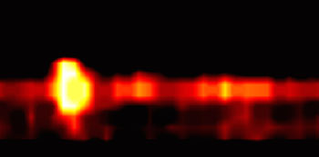
Image: Cancer cells expressing PSMA (Photo courtesy Kent Nastiuk / RPCI).
The researchers found that the PAI with an acoustic lens enabled good discrimination between cells with and without the PSMA-targeting agent attached with both imaging agents. They found however, that YC-27 fluoresced much brighter and that conjugation of the modified chromophores was more tractable. They concluded therefore that the urea-targeting agent represents a better approach to developing “louder” signals necessary to overcome the challenges of detecting PC by PAI in vivo. The study was published in the June 2016 issue of the Journal of Biomedical Optics.
“This proof-of-concept study demonstrates that this technology may allow for real-time monitoring of prostate cancer in patients during the course of active surveillance. For patients with more aggressive disease, the technology could offer more precise targeting of biopsies to confirm the need for definitive therapy,” said senior author Kent Nastiuk, PhD, assistant professor of cancer genetics and genitourinary cancers at RPCI. “This technology offers the potential to confirm the initial prostate cancer diagnosis, guide biopsies and monitor tumor volume — which is currently not measureable — for improved case management and treatment decision-making.”
PAI uses non-ionizing laser pulses delivered into biological tissues. Some of the delivered energy is absorbed and converted into heat, leading to transient thermoelastic expansion, and thus wideband ultrasonic emission, which can be detected by ultrasonic transducers and analyzed to produce images. The magnitude of the photoacoustic signal is proportional to the local energy deposition, which can be demonstrated by optical absorption contrast on the images of the targeted areas.
Related Links:
Roswell Park Cancer Institute
University of Rochester
Rochester Institute of Technology


