New Ultrasound Method Could Increase Understanding of Cancer Cells and Metastases
By MedImaging International staff writers
Posted on 01 Jun 2016
Researchers have discovered a new ultrasound method, called iso-acoustic focusing, that can be used to analyze and separate cells from blood.Posted on 01 Jun 2016
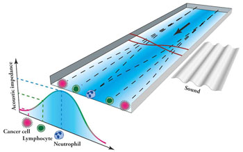
The new method developed at Lund University (Lund, Sweden) and Massachusetts Institute of Technology (MIT; Boston, MA, USA) exposes cells to ultrasound as they flow through a micro-channel inside a chip, and causes them to separate. The lateral movement of the cells enables the researchers to identify their acoustic properties, and could be used to detect the cell type, and distinguish between cancer cells of different origins.

Image: New ultrasound iso-acoustic technology could help increase awareness about the spread of cancer cells and metastases (Photo courtesy of Nature Communications / Lund University).
The study was published in the May 2016, issue of the online journal Nature Communications. The method could be used to measure the variation in the number of tumor cells in time and help determine whether medication can be effective in treatment or not.
Per Augustsson, Department of Biomedical Engineering, Lund University, said, “The vision is that our innovation will eventually be used in healthcare facilities, for example, to count and distinguish different types of cells in patients’ blood. It may seem odd that we are interested in the acoustic properties of blood cells and cancer cells. But we have been searching for new methods to separate cells in order to study them in more detail. Since we are looking for individual cells in a blood sample which contains billions of cells, the smallest overlap in size between the cancer cell and other blood cells will lead to thousands of blood cells ‘contaminating’ the cancer cells extracted through the separation. This is why we have now developed iso-acoustic focusing.”
Related Links:
Lund University
Massachusetts Institute of Technology


