Portable Ultrasound Helps Detect Fluid Retention
By MedImaging International staff writers
Posted on 26 May 2016
Nurses trained in the use of pocket ultrasound devices can accurately calculate fluid retention in cardiac patients and dispense diuretic drugs accordingly, according to a new study.Posted on 26 May 2016
Researchers at the Norwegian University of Science and Technology (NTNU, Trondheim, Norway) and Levanger Hospital (Norway) conducted a study to evaluate the clinical influence of focused ultrasound of the pleural cavities and inferior vena cava (IVC) as performed by specialized nurses to assess volume status in 62 patients who visited the heart failure (HF) outpatient clinic at Levanger Hospital on a total of 119 occasions.

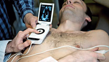
Image: A patient being examined with a portable ultrasound (Photo courtesy of the Research Council of Norway).
The HF outpatients underwent laboratory testing, anamnesis, and clinical examination by two nurses with and without an ultrasound examination of the pleural cavities and IVC using a pocket-size imaging device, in tandem with a cardiologist. The results showed that diuretic dosing differed between the two teams in 31 out of 119 consultations, meaning that in 89 of the paired consultations, the ultrasound examination changed treatment protocol. The study was published in the March 2016 issue of Heart.
“A pocket ultrasound device enables health personnel to detect signs of dehydration or worsening heart failure early, before the patient experiences symptoms of breathlessness, weight gain, and edema,” said lead author intensive care nurse Guri Gundersen. “Proper diuretic dose adjustment can quickly improve the patient's condition and prevent episodes of acute exacerbation of the disease that would require hospitalization.”
When the heart does not circulate blood normally, the kidneys receive less blood and filter less fluid out of the circulation into the urine. The extra fluid in the circulation congests in the lungs, the liver, around the eyes, and sometimes in the legs, and is the reason HF is also known as congestive heart failure.
Related Links:
Norwegian University of Science and Technology
Levanger Hospital


