Ultrasound Scanner Offers Enhanced View of Fetus
By MedImaging International staff writers
Posted on 24 Sep 2014
Using new ultrasound technology, clinicians can now visualize fetuses in the womb with unprecedented detail, allowing treatment that can be planned comprehensively before the baby is even born.Posted on 24 Sep 2014
The latest addition to the GE Healthcare (Chalfont St. Giles, UK) Voluson range of ultrasound scanners, the Voluson E10 system, features HDlive Silhouette and HDlive Flow applications that use ultrasound data in new ways to calculate depth, shape, and detail. Noise-removal, image enhancement, color, and light features are added, providing a final three-dimensional (3D) image. The versatile power of the system allows it to render these images in seconds, revealing what was once a grainy, grayscale, 2D image is now so clear that healthcare providers and patients can even “see a baby’s personality,” according to GE Healthcare spokespersons.

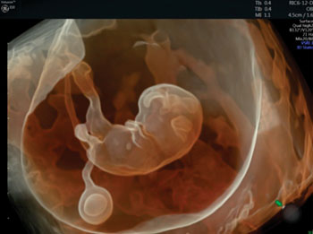
Image: Although clinically important, parents-to-be can be dismayed with the blurry gray image that appears with the first scan of their baby. This colored 3D image was captured using the Voluson E10 ultrasound system (Photo courtesy of GE healthcare).
A 3D scan is a still image of the baby in three dimensions. However, with 4D, the added dimension being time, the baby can be seen moving around in real time. HDlive technology adds a virtual light source to the image, calculating the location of shadows and even the translucency of the baby’s skin.
However, the image processing capabilities of the Voluson E10 can be used for more than superficial look at the fetus. The technology can be used to obtain images of the infant’s blood vessels, brain, heart, and other organs that show depth and structure in a way that helps provide the tiny details desired. This is particularly critical in the first trimester, where it is important to keep track of the baby’s growth.
Related Links:
GE Healthcare


