Photoacoustic Mammoscope Identifies Breast Cancer Using Light and Ultrasound
By MedImaging International staff writers
Posted on 06 Nov 2013
Routine screening can increase women’s chances of breast cancer survival by identifying the disease early and being able to treat it at this critical stage. A team of Dutch researchers developed a prototype of a new imaging tool that may soon help to detect breast cancer early, when it is at its most treatable stage.Posted on 06 Nov 2013
The new device, called a photoacoustic mammoscope, if effective would become a completely new strategy to image the breast and detecting cancer. In the place of X-rays, which are used in conventional mammography, the photoacoustic breast mammoscope uses a combination of ultrasound and infrared light to create a three-dimensional (3D) map of the breast. The researchers, from the University of Twente (Enschede, The Netherlands), published details of their device in an article published October 23, 2013, in The Optical Society’s (OSA) open-access journal Biomedical Optics Express.

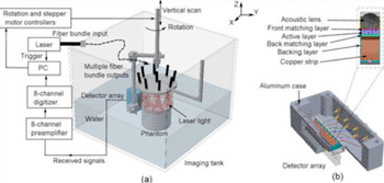
Image: A schematic illustration of the imaging system and the ultrasound detector (Photo courtesy of Wenfeng Xia, Biomedical Photonic Imaging group, University of Twente).
In the new technique, infrared light is delivered in billionth-of-a-second pulses to tissue, where it is scattered and absorbed. The high absorption of blood increases the temperature of blood vessels slightly, and this causes them to undergo a slight but rapid expansion. While indiscernible to the patient, this expansion generates identifiable ultrasound waves that are used to form a 3D map of the breast vasculature. Because cancer tumors have more blood vessels than the neighboring tissue, they are discernible in this image.
Currently the resolution of the images is not as fine as what can be captured with existing breast imaging techniques such X-ray mammography and magnetic resonance imaging (MRI). In future versions, Dr. Srirang Manohar, an assistant professor at the University of Twente, who led the research, Wenfeng Xia, a graduate student at the University of Twente, who was the first author on the new study, and their colleagues expect to enhance the resolution as well as add the capability to image using several different wavelengths of light at once, which is expected to improve detectability.
The Twente researchers have evaluated their prototype in the laboratory using phantoms (objects made of gels and other materials that mimic human tissue). In 2012, in a small clinical trial they revealed that an earlier version of the technology could successfully image breast cancer in women.
Dr. Manohar and his colleagues added that if the instrument were marketed, it would probably cost less than MRI and X-ray mammography tests. “We feel that the cost could be brought down to be not much more expensive than an ultrasound machine when it goes to industry,” said Dr. Xia.
The next phase of the research, the investigators report, will be to plan for larger clinical trials. Several existing technologies are already extensively used for breast cancer screening and diagnosis, including mammography, MRI, and ultrasound. Before becoming regularly used, the photoacoustic mammoscope would have to be at least as effective as those other techniques in large, multicenter clinical trials. “We are developing a clinical prototype that improves various aspects of the current version of the device,” said Dr. Manohar. “The final prototype will be ready for first clinical testing next year.”
Related Links:
University of Twente


