Color Flow Imaging Combined with Pulsed Wave Doppler to Create Ultrafast Doppler
By MedImaging International staff writers
Posted on 05 Sep 2011
Ultrasound imaging with pulsed wave Doppler provides a never-seen-before ultrahigh-frame rate of color flow clips that are up to 10 times faster than traditional color Doppler. Moreover, the same technology also acquires fully quantifiable Doppler data throughout the color box, enabling the generation of postprocessed pulsed wave Doppler spectra from multiple locations in the same image.Posted on 05 Sep 2011
SuperSonic Imagine (Aix-en-Provence, France), an ultrasound company that pioneered ShearWave elastography technology, announced another major breakthrough in ultrasound imaging: UltraFast Doppler. The company presented this new technology at the 2011 World Federation for Ultrasound in Medicine and Biology(WFUM) Euroson and Ultraschall meeting, held August 2011 in Vienna (Austria).

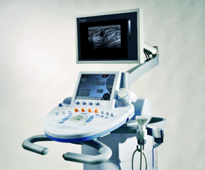
Image: The Aixplorer imaging system (Photo courtesy of SuperSonic Imagine).
Color flow imaging and pulsed wave Doppler are well-established ultrasound tools that analyze blood flow and are important for cardiovascular disease assessment and cancer diagnosis.
The Aixplorer’s UltraFast imaging platform, UltraFast Doppler, combines color flow imaging with pulsed wave Doppler. These new developments in Doppler technology introduce new era of flow imaging, workflow efficiency, and diagnostic effectiveness. Peter Burns, professor of medical biophysics at the University of Toronto (Canada) and senior scientist at Sunnybrook Health Sciences Center, Toronto, commented, “This technology promises to overcome two challenges faced by vascular sonographers every day when they use conventional duplex scanners. First, conventional color frame rates are too low to show real arterial hemodynamics, with pathology often masked by aliasing artifacts, and second, the pulsed Doppler examination is a separate and time-consuming process of searching for the optimal signal. UltraFast Doppler provides high frame rates, less aliasing, and the ability to show spectra at every point in a stored color loop. It provides more accurate flow imaging and will enable more effective workflow in the vascular lab.”
In 2008, SuperSonic Imagine introduced its Aixplorer MultiWave ultrasound system with excellent image quality and an elastography technique never before seen. The Aixplorer is the only system in the world that has ShearWave elastography imaging, the first major innovation in ultrasound since Doppler. This technology provides real-time, quantitative information about tissue stiffness by measuring and displaying local tissue elasticity on a color-coded map, in kilopascals. The evaluation of ShearWave elastography was confirmed in the largest breast clinical trial ever undertaken by an ultrasound company (1,800 lesions, 16 sites around the world) with validated reproducibility and increased ultrasound specificity while maintaining a high sensitivity. ShearWave elastography has been adopted by physicians worldwide as a tool to increase diagnostic precision and confidence.
“ShearWave elastography brought a new type of information to physicians to improve their diagnostic confidence. Today, with UltraFast Doppler, we are reinventing the Doppler analysis by breaking a 25-year-old rule in ultrasound of having to choose between flow imaging and flow quantification,” said Jeremy Bercoff, scientific expert and cofounder of SuperSonic Imagine. “We are very excited to introduce this new ultrafast technology feature and we are confident it will bring tremendous clinical benefits to physicians.”
SuperSonic Imagine is a multinational medical imaging company focused on developing the the Aixplorer ultrasound system. This system utilizes a unique MultiWave technology that enables the user to detect, characterize, and in the future, treat palpable and nonpalpable masses. MultiWave technology is based on combining two types of waves: an ultrasound wave that provides exceptional imaging in B-mode, and a shear wave, ShearWave elastography, which measures and displays the stiffness of tissue in kilopascals. ShearWave elastography is user-skill-independent with reproducible results. Aixplorer is CE Mark-approved since 2008, and has US Food and Drug Administration (FDA) clearance since 2009. The Aixplorer has several clinical applications: breast (and 3D breast), thyroid, abdomen, liver, musculoskeletal, prostate, and gynecologic (excluding obstetrics).
Related Links:
SuperSonic Imagine


