UltraSound System Guides Regional Anesthesia Blocks
By MedImaging International staff writers
Posted on 01 Sep 2010
An anesthesia-focused guidance system utilizes point-of-care ultrasound visualization for physician direction during peripheral nerve blocks.Posted on 01 Sep 2010
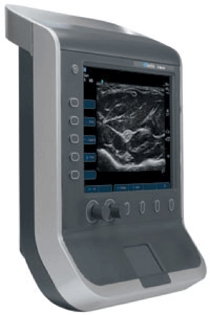
The SonoSite S-Nerve visualization tool helps regional anesthesiologists perform nerve block procedures in crowded, busy operating environments or anywhere in the hospital. With enhanced image quality, speed, and simplicity, the high-performance tool is designed to function exclusively as a guidance tool for regional nerve blocks and central line placement. A proprietary user interface, specialized software, and simple controls are matched to the processing power, image quality, and data management features of the Sonosite M-Turbo system; the enhanced processing power enables the simultaneous deployment of three advanced proprietary algorithms, SonoADAPT, SonoHD, and SonoMB, which combined produce dramatic improvements in image quality.

Image: The SonoSite S-Nerve visualization tool (photo courtesy SonoSite).
The S-Nerve tool can acquire the optimal image of hard-to-reach nerve structures with the turn of just two dials, and features a standard Video Electronics Standards Association (VESA) compliant mounting interface. When attached to the operating theater or block room wall or ceiling, the device has a zero footprint, a critical benefit in crowded environments. When mounted on an intravenous (IV) pole or hand-carried to the patient point-of-care, the device can easily be moved from patient to patient in a busy preoperative center.
The S-Nerve device is supplied with several curved and linear array transducers for virtually every anatomical and clinical anesthesia application, allowing the anesthesiologist to perform difficult upper and lower extremity blocks as well as confidently place intravascular lines in the desired vessels. The transducers, like the S-Nerve, are designed to withstand the demanding environment of the operating room or nerve block suite. They are also interchangeable with the SonoSite M-Turbo system and the new image optimization algorithms are available on all S-Nerve transducers and exam types. The SonoSite S-Nerve visualization tool is a product of SonoSite (Hitchin, United Kingdom).
"For the past six years, we have been actively encouraging ultrasound guidance for nerve blocks. This broad base of expertise is one of the prime reasons trainee anesthetists come to Derby, and straightforward controls of our S-Nerve systems are of great benefit to these inexperienced users,” said consultant anesthetist Adrian Searle, M.D., of Royal Derby Hospital (United Kingdom). "The robust nature of SonoSite's instruments is another advantage in this regard.”
Related Links:
SonoSite
Royal Derby Hospital


