Ultrasound Scan Improves Diagnosis of Heart Disease
By MedImaging International staff writers
Posted on 17 May 2010
New research shows that doing a simple ultrasound scan of the carotid artery significantly improves the prediction of heart disease, giving clinicians a better insight of who is at high risk for a heart attack. Posted on 17 May 2010
The new study, published in the April 7, 2010, issue of the Journal of the American College of Cardiology (JACC), revealed that approximately 23% of patients would be reclassified into a different risk group by adding information obtained from the noninvasive test and that risk prediction using this application was more accurate.

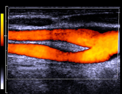
Image: Colored Doppler ultrasound scan of healthy blood flow through the junction of the common carotid artery (photo courtesy Zephyr / SPL).
"Today, up to 70% of people who have heart attacks are in a low or intermediate risk category for a heart attack when their risk is estimated using traditional risk prediction models. That's not very predictive, and we need to do better,” said Dr. Christie Ballantyne, director of the Center for Cardiovascular Disease Prevention at the Methodist DeBakey Heart & Vascular Center (Houston, TX, USA) and Baylor College of Medicine (Houston, TX, USA) and last author on the study. "Our research shows that a noninvasive ultrasound can give us a more complete snapshot of our patients' risk, so we can do a better job determining if they'll have a heart attack.”
This is significant because patients who are at higher risk could be treated more aggressively to prevent heart disease. Using ultrasound, researchers examined the carotid artery of 13,145 patients. The carotid artery feeds oxygenated blood from the heart to the brain. The researchers analyzed the thickness of the artery wall and the presence or absence of plaque inside the artery to determine if these factors influence risk for heart attack and coronary heart disease when added to traditional risk factors such as age, high blood pressure, high cholesterol, low good cholesterol, smoking, and obesity.
"We have known that people with heart disease tend to have thicker carotid arteries on ultrasound, but we now know how to use the artery thickness and presence or absence of plaque to better predict who is at risk for heart disease,” said Dr. Vijay Nambi, cardiologist with Methodist and Baylor, and first author on the study.
The analysis was performed using data from the Atherosclerosis Risk in Communities (ARIC) study. An online heart disease risk calculator is available at aricnews.net that will help doctors estimate risk, incorporating information about the carotid artery thickness and presence or absence of plaque.
Related Links:
Methodist DeBakey Heart & Vascular Center


