Ultra-High-Performance PET System Provides Never Before Seen Brain Images
Posted on 19 Jun 2024
Recent advancements in Positron Emission Tomography (PET) system technology have significantly enhanced their sensitivity, notably through improved time-of-flight capabilities. Yet, there has been only minimal enhancement in intrinsic resolution. Now, a new ultra-high-performance brain PET system enables direct measurement of brain nuclei as never before seen or quantified. This system, with its ultra-high sensitivity and resolution, delivers outstanding brain PET images that could drive breakthroughs in treating various brain disorders.
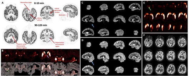
Designed by a collaborative team that included researchers from Yale University (New Haven, CT, USA), the NeuroEXPLORER PET scanner focuses on ultra-high sensitivity and resolution, as well as continuous head motion correction. In a comparative study, researchers performed human brain imaging using both the NeuroEXPLORER and the High Resolution Research Tomograph (HRRT), which was the previous state-of-the-art imaging tool. They administered multiple targeted radiopharmaceuticals to study synaptic density, dopamine receptors and transporters, muscarinic cholinergic receptors, and glutamate receptors. Subsequently, images from both scanners were analyzed and compared.

The results revealed a striking improvement in image contrast and quality with the NeuroEXPLORER as compared to the HRRT. The images produced by the NeuroEXPLORER displayed low noise and remarkable resolution, with clear focal uptake in specific brain nuclei. The research was presented at the 2024 Society of Nuclear Medicine and Molecular Imaging (SNMMI) Annual Meeting, and the grouping of images highlighting targeted tracer uptake in specific brain nuclei was selected as the 2024 SNMMI Henry N. Wagner, Jr., Image of the Year. The NeuroEXPLORER is poised to be a gamechanger in research for conditions such as Alzheimer’s disease, Parkinson’s disease, epilepsy, and various mental health disorders.
“The high resolution of NeuroEXPLORER images is due to the system’s unique detector design and exceptional sensitivity produced by its long axial field-of-view,” said Richard E. Carson, PhD, professor of Biomedical Engineering and of Radiology and Biomedical Imaging and Emeritus director of the PET Center at Yale University. “This technology will provide the opportunity for advanced research on all types of neuronal molecular and functional activity.”
Related Links:
Yale University


