Self-Powered Sensor to Make MRIs More Efficient
Posted on 03 Jun 2024
MRI scans are commonly used to diagnose a variety of conditions, anything from liver disease to brain tumors. During an MRI scan, a patient must stay entirely still for several minutes at a time, otherwise “motion artifacts” could appear and blur the final image. To ensure a clear picture, patient movement needs to be identified as soon as it happens, allowing the scan to stop and for the technician to take a new one. Motion tracking could be achieved using sensors embedded into the MRI table; however, magnetic materials can’t be used because metals interfere with the MRI technology itself. One technology that’s well-suited for this unique situation, and avoids the need for metal or magnetic components, is the triboelectric nanogenerator (TENG), which powers itself using static electricity generated by friction between polymers. Now, researchers have designed a TENG-based sensor that could be incorporated into an MRI machine to help prevent motion artifacts.
The self-powered sensor developed by researchers at the Chinese Academy of Sciences (Beijing, China) detects movement and shuts down an MRI scan in real time, improving the process for patients and technicians. The team created the TENG by sandwiching two layers of plastic film painted with graphite-based conductive ink around a central layer of silicone. These materials were specifically chosen as they would not interfere with an MRI scan. When pressed together, electrostatic charges from the plastic film moved to the conductive ink, creating a current that could then flow out through a wire.
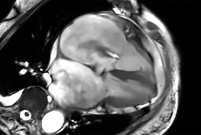
 attached to a headrest within an MRI machine detects patient movement in real time (Photo courtesy of ACS Sensors 2024, DOI: 10.1021/acssensors.4c00319)")
This sensor was incorporated into an MRI table designed to lay under a patient’s head. In tests, when a person turned their head from side to side or raised it off the table, the sensor detected these movements and transmitted a signal to a computer. Then, an audible alert played, a pop-up window on the technician’s computer appeared and the MRI scan ceased. The researchers say that this work could help make MRI scans more efficient and less frustrating for patients and technicians alike by producing better images during a single procedure.
Related Links:
Chinese Academy of Sciences