New Reporting Style Improves Accuracy and Speed of Reading Radiology Scans
Posted on 06 Dec 2023
Certain health issues, such as calcified arteries, infections, minor bone fractures, or cancerous tumors, often remain hidden within our bodies. Special imaging techniques like X-rays, MRIs, or CT scans are crucial for detecting these issues. Interpreting the bluish, black and white images produced by these scans requires the expert eye of a radiologist, who is adept at identifying anomalies in images of our brains, lungs, arteries, bones, and muscles. Radiologists specialize in spotting irregularities and generate detailed reports for physicians to confirm diagnoses and inform treatment plans. Radiologists typically review a vast range of images, from about 200 in simpler cases like X-rays to 20,000 in more intricate MRI scans, leading to an average of 50 to 60 reports each day.
Technological advancements such as AI-powered speech-to-text conversion tools, can assist radiologists in dictating and completing important reports for the patient’s healthcare team, although there can be mistakes in the transcription that can have ripple effects. A research team at Florida International University (Miami, FL, USA) explored the effectiveness of a new reporting style that is more concise yet remains structured and easy to comprehend. This style leverages voice command technology for dictation, focusing solely on documenting abnormal findings that radiologists are trained to identify and that are critical for doctors' awareness. Essentially, the radiologists verbalize only the irregularities they observe. This approach was found to be more efficient than the traditional checklist-style reporting commonly utilized in radiology.

The standard checklist reporting style often requires radiologists to divide their attention between two screens - one showing the diagnostic images and the other displaying the transcribed spoken findings. This process can be cumbersome and distracting, particularly when correcting errors arising from voice recognition software misinterpreting spoken words. In contrast, the new method allows radiologists to concentrate fully on the images, mentally going through the checklist based on their training. They only dictate the abnormal findings, reviewing the report for accuracy after completing their interpretation of the images.
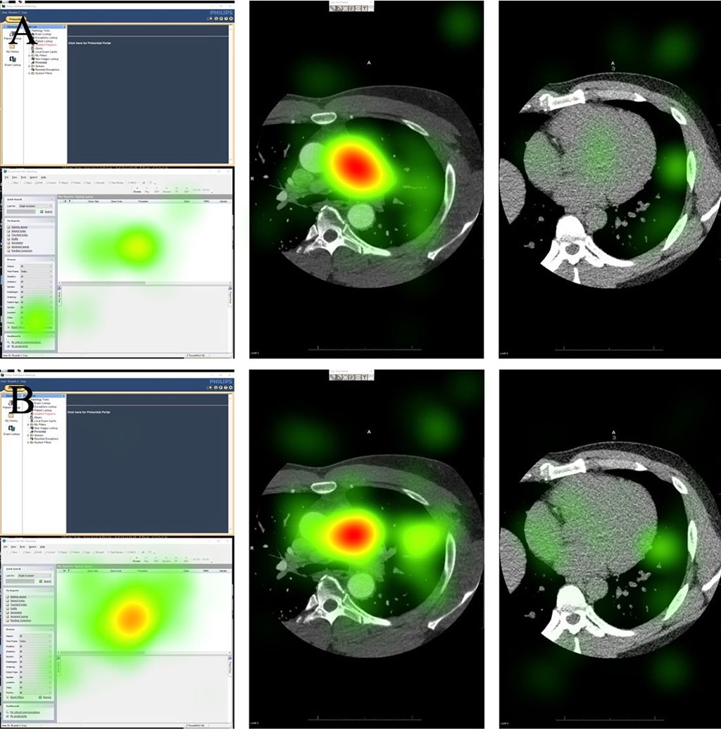
In the new study, experienced, board-certified radiologists used eye-tracking goggles to review a variety of X-rays, MRIs, and CT scans, totaling over 150 studies — 76 with the new dictation style and 81 with the standard checklist. This new approach not only enhanced the radiologists' focus on the diagnostic images but also reduced inaccuracies and dictation time, benefiting both patients, who rely on accurate reports for their treatment, and overburdened radiologists. The new dictation method, using voice commands, cut the average dictation time by about half without affecting the total time taken for interpretation or examination. The researchers view this study as a foundation for future advancements, anticipating that evolving conversational and generative AI technologies could further enhance the efficiency and accuracy of radiology reporting.
“I saw firsthand how this new dictation process significantly enhanced radiologists’ focus on the diagnostic images,” said Mona Roshan, an FIU third-year medical student and the study’s first author. “The eye-tracking software validated our results, because it allowed us to create heatmaps showing that radiologists were directing their attention to the imaging. This is better for the patient, as a heightened focus on the images means that radiologists are dedicating more time to interpreting potential abnormal findings, ultimately improving patient care.”
Related Links:
Florida International University


 Guided Devices.jpg)


