AI X-Ray Tool Estimates Bone Mineral Density for Early Diagnosis of Osteoporosis
Posted on 13 Oct 2023
Osteoporosis is a common health issue that leads to low bone mineral density (BMD), making bones fragile and increasing the risk of fractures. Diagnosing this condition typically involves specialized and often costly tests like dual-energy X-ray absorptiometry (DXA) and quantitative computed tomography (QCT). Because of these limitations, there's a need for more convenient and budget-friendly screening options. Recently, machine learning techniques that use X-ray images to estimate BMD have become more popular, but these often require extensive training data. Researchers have now come up with a machine learning method that offers a simpler way to screen for osteoporosis and other bone conditions early on.
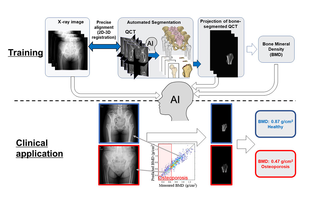
Researchers at Nara Institute of Science and Technology (NAIST, Nara, Japan) have devised an innovative method that utilizes a type of machine learning known as the hierarchical learning framework. This method estimates BMD from standard X-ray images. The research team used original QCT scans from patients to create a virtual X-ray image of the bone area, aligning it precisely with actual patient X-rays. This data was then used in three distinct training phases to develop a final BMD estimation model. Initially, the model focused on breaking down X-ray images to create a virtual X-ray image of the bone area. In the final phase, the model was trained to recognize the relationship between these virtual X-ray images and BMD values.

This method was able to accurately estimate BMD using just a single X-ray image and demonstrated high effectiveness even with a couple of hundred datasets of CT and X-ray image pairs. The model not only provides the BMD value but also generates a virtual X-ray image that shows the distribution of bone density, making the results easier to understand. To assess its effectiveness compared to traditional methods like DXA and QCT, the researchers performed validation tests with real clinical data. The BMD values obtained through this new method showed a strong correlation with those derived from DXA and QCT, confirming its reliability.
Additional validation tests further demonstrated the robustness of this method. It produced consistent BMD estimates despite changes in patient positioning or different levels of image compression. The outcomes indicate that this new method has enormous potential for regular medical use. It offers a way to conveniently screen for osteoporosis and monitor treatment, enabling timely intervention and potentially improving the lives of those living with the condition.
"Osteoporosis is generally diagnosed at advanced stages once its symptoms become apparent. X-ray images can be valuable for opportunistic diagnosis, but efficiently extracting BMD information from these has been a significant challenge,” said Yoshito Otake from NAIST. “We hoped to solve this problem by using information derived from the computed tomography (CT) image in the training stage to develop a model for an accurate, efficient, and explainable BMD estimation solely from an X-ray image."
Related Links:
NAIST


