3D-Printed Marker Optimizes Portable Radiography
By MedImaging International staff writers
Posted on 17 Jun 2021
A novel head-of-bed (HOB) angulation device significantly improves the diagnostic performance of portable chest and abdominal x-rays. Posted on 17 Jun 2021
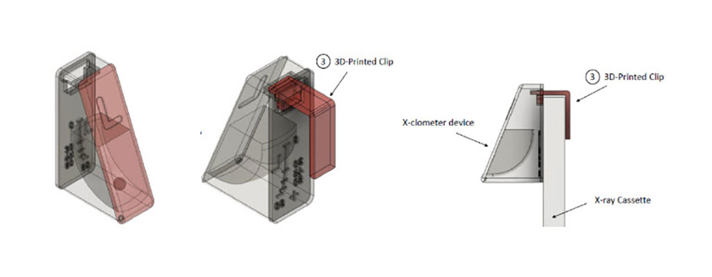
Developed at the U.S. National Institutes of Health (NIH; Bethesda, MD, USA), the X-clometer is used to resolve the location the x-ray cassette, relative to the x-ray source, and divergent x-rays. The device, which is placed in the upper right corner of the field of view (FOV), is essentially a left or right marker that also quantifies the HOB angle from supine (0 degrees) to upright (90 degrees). The approximate angle is determined by a ball bearing that rolls freely within the curved passageway of the device to indicate the angle of the patient, cassette, and x-ray tube during the chest x-ray.

Image: The X-clometer resolves relative angulation of an x-ray (Photo courtesy of NIH)
The patented technology improves performance of portable chest and abdominal x-rays, and allows reliable comparisons of patient condition over time and improved care for intensive care unit (ICU) patients. For example, the need for immediate drainage resulting from pleural effusion can be more effectively assessed. The X-clometer was presented at the Society for Imaging Informatics in Medicine (SIIM) annual meeting, held online during May 2021.
“We believe that knowing the degree of inclination across serial exams will help negate the need to bring patients to the department with numerous chest drains, IV lines, and other support devices,” said device presenter Raisa Freidlin, DSc, of the NIH. “In addition, following further evaluation and actual use, X-clometer may decrease the need for obtaining CT scans, which would reduce unnecessary radiation exposure and additional expenses.”
The system was created using the 3D model assembly programs Solidworks and Fusion 360. Using 3D computer-aided detection software, the researchers recently improved reading accuracy in the 60- to 90-degree range, which also optimized size and positioning of the device. The X-clometer 3D print files are publicly available on the NIH's 3D Print Exchange website.
Related Links:
U.S. National Institutes of Health
NIH's 3D Print Exchange


